
 Türkçe
 Deutsch
 العربية

Pediatric urology is a medical specialty that covers the diagnosis, treatment, and follow-up of urinary tract and reproductive system diseases in individuals from infancy to adolescence. Congenital anomalies and functional disorders constitute the main subjects of examination in this field.
Among pediatric urology diseases, congenital problems such as vesicoureteral reflux, hydronephrosis, and undescended testis are common, and early diagnosis is of great importance. Since the progression of these diseases may affect kidney functions, regular follow-up and appropriate treatment planning are necessary.
Urinary tract infections and voiding disorders in children are among the important clinical topics of pediatric urology, and recurrent infections may be an indication of underlying structural problems. Ultrasonography and advanced imaging methods are used in the diagnostic process.
Pediatric urology treatment methods include surgical and medical approaches, and minimally invasive techniques are currently at the forefront. Laparoscopic surgery and endoscopic interventions accelerate the patient’s recovery process and reduce the risks of complications.
| Things You Should Know | Information |
| Definition | Pediatric urology is a subspecialty of urology specific to children that deals with the diagnosis and treatment of congenital or acquired urinary and genital system disorders. |
| Age Group | It covers children from newborns up to 18 years of age. |
| Main Diseases | Hypospadias, vesicoureteral reflux (VUR), hydronephrosis, undescended testis, posterior urethral valve (PUV), urinary tract infections (UTI), bladder dysfunction, phimosis, varicocele. |
| Symptoms | Frequent urination, urinary incontinence, pain while urinating, non-retractable foreskin, swelling in the scrotum, recurrent urinary tract infections. |
| Diagnostic Methods | Physical examination, ultrasonography, voiding cystourethrography (VCUG), scintigraphy (DMSA), urodynamics, MRI or CT imaging. |
| Treatment Options | Drug treatment, surgical intervention (for example hypospadias repair), bladder training, endoscopic interventions, laparoscopic surgery. |
| Conditions Requiring Surgery | Hypospadias, undescended testis, phimosis, posterior urethral valve, severe VUR or hydronephrosis cases. |
| Multidisciplinary Approach | Collaboration with different disciplines such as nephrology, radiology, pediatric surgery, and physiotherapy may be required. |
| Follow-up Process | Long-term follow-up may be required; monitoring kidney functions, evaluating growth and development, and preventing recurrent infections are important. |
| Recommendations for the Family | It is important to notice symptoms early, attend regular doctor check-ups, and follow medication and hygiene recommendations. |

Prof. Dr. Berkan Reşorlu
Urology Specialist
Urology Specialist
Prof. Dr. Berkan Reşorlu, born in Ankara in 1979, completed his medical education at Ankara University between 1997-2003 and his urology specialization at Ankara University Faculty of Medicine between 2003-2008. Academically, he received the title of associate professor in 2013 and professor in 2022.
He has carried out extensive series studies in the field of endoscopic stone surgery, introduced surgeries applied to children under 1 year of age into the literature, and gained international recognition with his stone scoring system and experimental surgical models.


What Is Hypospadias and How Does the Surgical Treatment Process Work?
Hypospadias, commonly known among the public as prophet circumcision or being born half-circumcised, is a condition in boys in which the urinary opening is located at any point on the underside of the penis instead of its natural place at the tip of the penis. This condition, encountered in approximately one out of every 250 to 300 live male births, is a very important issue that requires mastery of anatomical details and a high level of surgical experience. A complex interaction of hormonal imbalances in the womb, genetic predispositions, and certain environmental factors plays a role in its formation. Although this condition naturally causes great concern in families, it can be treated quite successfully with today’s medical possibilities.
In the clinical evaluation of a child with hypospadias, the location of the urinary opening alone does not carry meaning by itself. Many factors such as the general structure of the penis, the tissue quality of the urinary canal, and the distribution of the foreskin are considered as a whole. In particular, penile curvature, which is seen in a quarter of the cases and causes the penis to bend downward, is one of the greatest obstacles to surgical success and must definitely be corrected during surgery. If the urinary opening is located very close to the scrotum, different anatomical problems such as undescended testis or inguinal hernia may also accompany this condition.
The main anatomical structures evaluated before surgery are:
- Location of the urinary opening
- Amount of foreskin
- Penile curvature
- Quality of the urinary canal
- Condition of the testes
The most ideal accepted time interval for surgical intervention is between 6 and 18 months. There are very valid physical and psychological reasons for preferring this period. During infancy, cell renewal and tissue healing speed are very high. In addition, since the baby still uses diapers, wound care and protection from infection after surgery are much easier to manage. Most importantly, because the child has not yet completed psychosexual development and is not aware of the genital region, this surgical process is overcome without experiencing a trauma that will be remembered later in life. In some cases, the penis size may not be large enough for surgical intervention. In such cases, the chances of surgical success are increased by improving tissue quality and penis size with special hormone creams applied externally a few months before surgery.
The surgical techniques used in hypospadias repair are:
- Tubularized incised plate method
- Onlay flap method
- Meatal advancement method
- Two-stage graft repair
Which of these methods will be chosen depends entirely on the position of the opening and the quality of the tissue. In cases where the urinary opening is close to the tip of the penis, a new tube is created using the existing tissue, while in more proximal cases, a completely new urinary canal is constructed with grafts taken from the foreskin or inside the mouth. After these surgeries, problems such as urine leaking from a different place or narrowing in the canal may sometimes occur, but these are also conditions that can be resolved with additional interventions.
What Causes the Problem of Undescended Testis and When Should Intervention Be Performed?
Undescended testis stands out as one of the most common surgical problems in boys. While its incidence is relatively low in full-term babies, this rate reaches much higher levels in premature babies born early. During the developmental process in the womb, the testes begin to form just below the kidneys and gradually descend downward to settle into the scrotum months before birth. The picture of undescended testis emerges as a result of this journey being interrupted due to hormonal deficiencies or anatomical obstacles.
The testicular conditions detected during physical examination are:
- Palpable testis
- Non-palpable testis
- Retractile testis
- Ectopic testis
In a great majority of cases, the testis is stuck somewhere in the inguinal canal and can easily be felt by hand during the doctor’s examination. However, in some cases, the testis may remain deep in the abdominal cavity or may never have developed congenitally. In such non-palpable testis cases, imaging methods such as ultrasound or MRI are generally insufficient. At this point, the most definitive and reliable diagnostic method is laparoscopy, in which the inside of the abdomen is examined with the help of a small camera. Thanks to laparoscopy, the location of the testis is identified and surgery can be performed in the same session. In the condition called retractile testis, the testis is actually in the scrotum, but due to an excessive reflex of the muscles, it frequently moves upward. If the testis is seen in the scrotum during a warm bath or while the child is sleeping, this condition usually does not require surgical intervention and is only followed at regular intervals until puberty.
Timing is of vital importance in the treatment of undescended testis. The internal temperature of the scrotum is a few degrees lower than body temperature, and the testes need this cool environment to develop healthily and produce sperm. If the testis remains in the abdomen or groin, that is, in a warm environment, for a long time, the reproductive cells inside it begin to suffer irreversible damage from the first birthday onward. Therefore, current medical approaches foresee that surgery should be performed from the sixth month onward and definitely not be left after the eighteenth month. In delayed cases, the risk of infertility in adulthood can reach serious levels. In addition, the risk of developing cancer at later ages in undescended testes is much higher than in a testis in its normal position. Although surgery performed at an early age does not completely eliminate this risk, bringing the testis down into the scrotum makes it possible for a possible mass to be noticed by hand early and treated.
Contact us now to get detailed information about treatments and to make an appointment!
What Are the Diagnostic Methods in the Case of Kidney Reflux (Vesicoureteral Reflux)?
Vesicoureteral reflux, or kidney reflux as it is commonly known among the public, is the condition in which urine that should normally only be expelled outward after collecting in the bladder flows backward through the urinary channels toward the kidneys. In a healthy urinary tract, there is a thin one-way valve mechanism at the point where the urinary channel enters the bladder. Thanks to this valve, urine cannot go backward when the bladder contracts. However, when this valve becomes unable to perform its function due to a congenital structural weakness or other diseases that excessively increase the pressure inside the bladder, reflux disease begins. The urine that escapes backward causes high pressure inside the kidney, and if there are bacteria in this urine, it leaves permanent inflammatory damage in the kidney tissue.
The radiological stages determining the severity of the disease are:
- First degree
- Second degree
- Third degree
- Fourth degree
- Fifth degree
Standard ultrasonography is generally insufficient in the diagnosis of the disease and in determining its degree. This is because although ultrasound shows the enlargement in the kidney, it cannot clearly show whether urine is momentarily flowing backward. Therefore, for a definitive diagnosis, a special radiological method is used in which a thin catheter is inserted into the bladder, contrast fluid is administered inside, and images are taken while the child urinates. In this method, situations in which urine only escapes into the channel are called first degree, while situations in which it reaches the kidney but does not cause enlargement are called second degree. The most severe picture, in which urine causes excessive enlargement in the renal pelvis, twisting in the channels, and thinning in the kidney tissue, is classified as fifth degree. In addition, to understand how much the kidneys have been damaged by this backward flow and whether there is any loss of function, kidney scintigraphy with nuclear medicine methods must definitely be performed.
What Treatment Options Are Available for Kidney Reflux (VUR)?
The treatment of kidney reflux is planned entirely on an individual basis according to the degree of the disease, the child’s age, and the frequency of febrile urinary tract infections experienced. In low-grade reflux, the possibility of the disease resolving spontaneously is quite high as the child grows, thanks to the development of the bladder muscles and the maturation of the anatomical structure. During this waiting process, protecting the kidneys from possible infections is of vital importance. For this purpose, long-term, very low-dose prophylactic antibiotic treatment is started for the child and a strict follow-up program is carried out with regular urine tests. However, if the child continues to have frequent febrile infections despite the use of prophylactic antibiotics, if new areas of damage are detected on kidney scintigraphy, or if the degree of reflux is too high to resolve spontaneously, surgical intervention options come into play.
The main surgical treatment methods applied are:
- Endoscopic injection
- Open surgical reimplantation
- Laparoscopic reimplantation
- Robotic surgical reimplantation
Today, the first choice in the treatment of low- and moderate-grade reflux is generally endoscopic injection, which is a closed method. In this method, no incision is made in the body. Special camera-equipped instruments are inserted through the urinary opening, and a special tissue-compatible filler material is injected into the lower part of the channel through which urine flows back to the kidney. This filler material creates a small mound in that area and enables the valve mechanism to function again. The procedure is very short and the child can return home the same day.
However, in severe fourth- and fifth-degree reflux where the injection method is insufficient, open or robotic surgical methods are used. In this major surgical procedure, the urinary channel is carefully separated from the place where it is connected to the bladder, and the channel is sutured there again by creating a longer, completely new tunnel within the bladder muscle. Thanks to this new tunnel, the mouth of the channel closes when the bladder fills or contracts, and the backward flow of urine is permanently prevented. The success rate of these surgeries is very close to one hundred percent and they provide definitive protection for the kidneys.
How Is Ureteropelvic Junction (UPJ) Obstruction Understood and What Are Its Surgical Solutions?
Ureteropelvic junction obstruction, or UPJ obstruction for short, is a blockage that occurs exactly at the junction point between the pelvic part where the kidney filters and collects urine and the thin channel that carries urine to the bladder. We can compare this to a blockage right at the mouth of a sink drain pipe. Water flows and accumulates in the sink, but cannot easily go down. Since the urine continuously produced in the kidney cannot pass through this narrow bottleneck, it collects in the renal pelvis and over time causes the kidney to swell, that is, hydronephrosis. This condition, which is the most common cause of kidney enlargement in newborn babies, is usually noticed incidentally during routine pregnancy ultrasounds performed while the baby is still in the womb.
The tests used during the diagnosis and follow-up stage are:
- Ultrasonography
- Complete urinalysis
- Diuretic renography
- Magnetic resonance imaging
In the first weeks after the baby is born, ultrasound is repeated and the size of the swelling in the kidney is measured. However, ultrasound cannot indicate how serious the blockage is or how much it impairs kidney function. For this, a very special test called diuretic renography is performed in the nuclear medicine department. Thanks to this test, it is measured what percentage both the right and left kidney contribute to total kidney function, and it is calculated in how many minutes the urine descends from the obstruction point. If there is a significant decrease in the kidney’s functional capacity or if the urine drainage time has risen far above critical levels, surgery is then decided upon to save the kidney.
The surgical techniques preferred for this operation are:
- Open pyeloplasty
- Laparoscopic pyeloplasty
- Robotic pyeloplasty
The main purpose of the surgery is to completely cut out and remove the diseased section of the channel that is narrow and blocks the passage of urine, and to re-suture the remaining healthy renal pelvis and urinary channel to each other with a much wider opening. Instead of open surgeries that used to be performed with a large flank incision, laparoscopic or robotic surgeries performed today by entering through much smaller holes are preferred. Especially in cases where an extra vessel going to the lower part of the kidney (crossing vessel) causes obstruction by pressing on the channel from the outside, the three-dimensional, magnified view and high maneuverability provided by robotic surgery offer great advantages to the surgeon. The vessel is preserved, the urinary tract is moved to the front of the vessel, and the obstruction is removed. Thanks to these closed methods that we call minimally invasive, the child has much less pain after surgery and the hospital stay is shortened.
Contact us now to get detailed information about treatments and to make an appointment!
In Which Situations Is Circumcision Necessary and How Is Faulty Circumcision Corrected?
Circumcision, medically called circumcision, is the surgical removal of the skin covering the tip of the penis in boys. Beyond being only a religious or cultural ritual, it is a medical intervention with very serious protective effects on child health when performed under proper conditions and by experienced hands. It is known in particular that circumcision performed in the neonatal period, that is, within the first month of life, reduces the risk of urinary tract infections, which can be very dangerous during that period, by approximately tenfold. In addition, its protection against penile cancer in the long term and its hygienic advantages have also been scientifically proven.
However, the perception of circumcision in our society as a very simple and ordinary procedure unfortunately causes it to be performed in inappropriate environments by unqualified people, resulting in complications that are very difficult to remedy. While cutting too much of the foreskin hinders the development of the penis, cutting too little prepares the ground for infections. Accidental cutting of the urinary channel is a condition that requires much more extensive surgeries.
The most common problems encountered after faulty circumcision are:
- Skin adhesions
- Narrowing of the urinary opening
- Buried penis
- Skin bridges
Among these problems, the condition of buried penis in particular requires separate attention. It is the condition in which, due to the child being overweight or because of anatomical structure, the penis becomes buried into the fatty tissue in the groin area and becomes invisible from the outside. Families usually bring their children to the doctor for a second circumcision thinking that the circumcision was incomplete or that the skin has grown back. However, these children should definitely not undergo a repeat classical circumcision. A mistakenly performed second incision causes the penis to become completely trapped under the skin. In cases of buried penis, much more complex penoscrotal revision surgeries are needed, in which the ligaments holding the penis at its root and pulling it downward are released and the penis is fixed outward. In addition, narrowing that may be seen in the urinary opening after circumcision causes complaints of urinating through a thin stream and straining. This condition can be treated easily with a very short outpatient intervention called meatotomy.
What Are the Differences Between Hydrocele and Inguinal Hernia?
Hydrocele and inguinal hernia seen in children are two different reflections of the same anatomical defect that occurs in the last months of the developmental process in the womb. While the baby is in the womb, as the testes descend from inside the abdomen toward the scrotum, they also pull a small tube from the peritoneal layer downward with them. Close to birth, this tube is expected to close completely and disappear. If this channel remains open, a connection continues between the inside of the abdomen and the scrotum.
The main points of distinction between these two conditions are:
- Difference in content
- Risk of strangulation
- Timing of intervention
- Possibility of spontaneous healing
If the channel that remains open is very narrow, the organs inside the abdomen cannot pass through it, and only the intra-abdominal fluid leaks downward drop by drop and accumulates around the testis. This condition is called hydrocele. The swelling usually becomes prominent when the child is standing, straining, or crying, and when sleeping, the scrotum becomes smaller because the fluid returns to the abdomen. Since it contains only fluid and does not carry the risk of organ entrapment, it is generally not a very urgent condition. Considering the possibility that the channel may close on its own, it can be observed without surgery until the age of two.
However, if the channel remains widely open, the intestines inside the abdomen or, in girls, the ovaries may protrude downward through this channel. This is called inguinal hernia. The entrapment of the descended intestinal loops in this narrow channel may impair blood flow and in a very short time cause the tissue to decay, creating a life-threatening situation. This picture is called strangulated hernia. Because of this high risk, once inguinal hernia is diagnosed in a child, it is essential that the channel be surgically tied and closed as soon as possible without waiting for age or weight.
What Are the Main Causes and Treatment of Bedwetting (Enuresis) in Children?
Bedwetting at night, medically termed nocturnal enuresis, which is one of the most distressing problems of childhood and one that wears families out the most, is the condition in which the child continues to wet the bed involuntarily during sleep despite having completed five years of age. This problem is much more common than thought; it is seen in one out of every ten seven-year-old children. As age progresses, it tends to improve spontaneously at a certain rate every year with the maturation of the nervous system. However, considering the loss of self-confidence, withdrawal, and family tensions that the child may experience during this process, the condition must definitely be managed with professional support.
Cases of enuresis are basically divided into two groups. If the child only wets the bed at night and has no leakage or urgency problem during the day, this condition is monosymptomatic. However, if daytime dribbling, sudden urgency causing leg crossing, frequent or very infrequent urination, and especially constipation accompany nighttime wetting, this condition is considered complicated and requires a much more detailed examination.
The voiding and fluid intake diary kept by families during the diagnostic process provides very valuable information. A simple but effective formula is used to understand whether the child’s bladder is of an age-appropriate size. Bladder capacities that remain far below this expected value indicate that the bladder has not grown enough or is overactive.
The main approaches used in the treatment process are:
- Lifestyle changes
- Urotherapy applications
- Use of an alarm device
- Drug treatments
It is generally preferred to start treatment during the period when the child begins primary school. The first step is always the lifestyle adjustments we call urotherapy. The child’s water consumption during the day is balanced, fluid intake is restricted in the evening hours, carbonated and caffeinated drinks are prohibited, and most importantly, the child is made to go to the toilet at regular intervals during the day. If there is constipation, it must definitely be treated simultaneously, because a full large intestine narrows bladder capacity by putting pressure on the bladder.
Blog Posts

Genital Warts: Which Doctor or Department Should You See?
When genital warts are noticed, the [...]
Read More
13
Jul
Jul

Urinary Tract Infection (UTI): Which Doctor or Department Should You Visit?
The department patients experiencing urinary tract [...]
Read More
13
Jul
Jul

Kidney Stones: Which Doctor or Specialist Should You See?
The primary specialty that patients with [...]
Read More
13
Jul
Jul
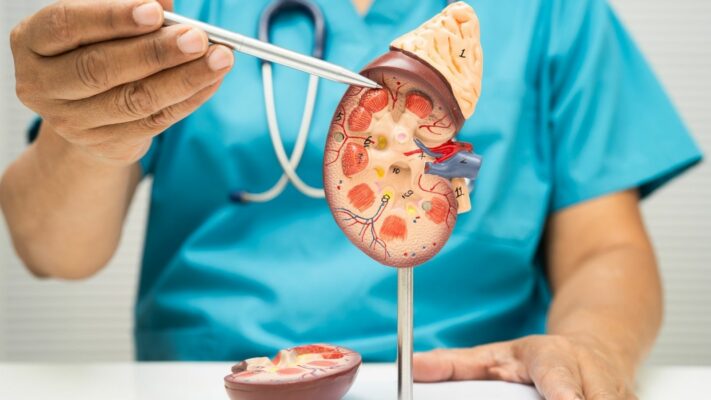
Kidney Diseases: Which Doctor or Department Should You See?
The primary medical specialty that patients [...]
Read More
13
Jul
Jul

Sexually Transmitted Diseases (STDs): Which Doctor Should You See?
The answer to the question of [...]
Read More
13
Jul
Jul

Kidney Cysts: Which Doctor or Department Should You See?
The primary medical specialties that patients [...]
Read More
13
Jul
Jul
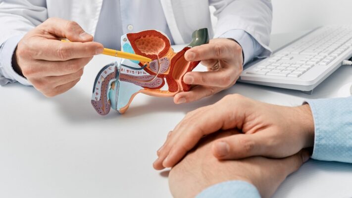
Prostate Problems: Which Doctor or Specialist Should You See?
The medical specialty that men experiencing [...]
Read More
13
Jul
Jul

Premature Ejaculation: Which Doctor Should You See for Treatment?
The first and primary medical specialty [...]
Read More
13
Jul
Jul