
 Türkçe
 Deutsch
 العربية

Kidney stones are hard structures formed as a result of the crystallization of minerals and salts in the urinary tract and usually present clinically with severe pain, hematuria, and impaired urine flow. Early diagnosis and appropriate treatment are of critical importance in preventing complications.
The causes of kidney stone formation develop depending on factors such as low fluid intake, diets high in oxalate and sodium, and genetic predisposition. Metabolic disorders and urinary tract infections are also among the important factors that increase the risk of stone formation.
The symptoms of kidney stones appear as sudden flank pain, nausea, vomiting, and blood in the urine. The severity of symptoms varies depending on the size and location of the stone, and in some cases it may follow an asymptomatic course.
Kidney stone treatment methods include medical treatment, extracorporeal shock wave lithotripsy, or surgical interventions depending on the size of the stone and the patient’s clinical condition. In addition, adequate fluid intake and dietary adjustments are effective in reducing the risk of recurrence.
| What You Need to Know | Information |
| Definition | Kidney stone (nephrolithiasis) is the condition in which minerals and salts found in urine crystallize and form hard structures inside the kidney or in the urinary tract. |
| Formation Mechanism | Crystals form as a result of an increase in the concentration of substances such as calcium, oxalate, and uric acid in the urine and insufficient fluid intake, and over time they combine and become stones. |
| Stone Types | Calcium oxalate stones are the most common. In addition, calcium phosphate, uric acid, struvite (infection stones), and cystine stones may also form. |
| Risk Factors | Insufficient fluid intake, a diet high in salt and protein, family history, obesity, recurrent urinary tract infections, some metabolic diseases, and living in a hot climate are among the risk factors. |
| Symptoms | Severe flank pain (renal colic), pain radiating to the groin, blood in the urine (hematuria), nausea, vomiting, frequent urination, burning during urination, and sometimes fever may be seen. |
| Pain Characteristic | The pain usually starts suddenly, increases and decreases in waves. It often starts on the side of the waist and spreads toward the groin area. |
| Diagnostic Methods | Urinalysis, blood tests, ultrasonography, computed tomography (CT), and direct urinary system radiography are used in diagnosis. |
| Treatment – Small Stones | Stones smaller than 5 mm can usually pass spontaneously with plenty of fluid intake, painkillers, and in some cases medications that facilitate stone passage. |
| Treatment – Large Stones | In large or non-passing stones, extracorporeal shock wave lithotripsy (ESWL), ureteroscopy, percutaneous nephrolithotomy, or rarely surgical intervention may be applied. |
| Complications | Urinary tract obstruction, infection, loss of kidney function, and recurrent stone formation may develop. |
| Prevention Methods | Adequate daily fluid intake (enough to give the urine a light color), restriction of salt and excessive animal protein, balanced consumption of oxalate-rich foods, and regular medical follow-up are recommended. |
| Risk of Recurrence | The risk of recurrence is high in individuals with kidney stones. Therefore, metabolic evaluation and lifestyle changes are important. |

Prof. Dr. Berkan Reşorlu
Urology Specialist
Urology Specialist
Prof. Dr. Berkan Reşorlu, born in Ankara in 1979, completed his medical education at Ankara University between 1997-2003 and his urology specialization at Ankara University Faculty of Medicine between 2003-2008. Academically, he received the title of associate professor in 2013 and professor in 2022.
He has carried out extensive series studies in the field of endoscopic stone surgery, introduced surgeries applied to children under 1 year of age into the literature, and gained international recognition with his stone scoring system and experimental surgical models.


What is a kidney stone and how does it form in our body?
The kidneys are vital organs that continuously work like a treatment facility, filtering waste substances in the bloodstream and removing them from the body through urine. Under normal conditions, these waste minerals are dissolved in liquid form within the urine. Sometimes, due to a significant decrease in fluid intake or a metabolic problem, the amount of water in the urine decreases and the concentration of minerals that need to be excreted rises far above normal. This condition is called supersaturation of the urine.
These concentrated minerals, especially substances such as calcium, oxalate, uric acid, or phosphate, attach to the delicate tissues on the inner surface of the kidney and form microscopic crystals. Over time, these crystals stick together, grow layer by layer, and gradually harden, forming the calcified structures we call kidney stones. The formation process is usually extremely silent; patients may live for months or even years completely unaware of a stone growing inside. These stones may be as small as a grain of sand that is difficult to see with the naked eye, or they may reach enormous and complex sizes large enough to completely fill all the inner chambers of the kidney where urine is collected.
Who is more prone to developing kidney stones and what are the main risk factors?
It is a known and statistically proven fact that some people in society are much more prone to this condition than others. The individual’s anatomical kidney structure, the temperature conditions of the geography they live in, their occupation, and most importantly their daily dietary habits play a very major role in this process. The risk is much higher especially in individuals living in very hot climates, performing physical exertion under the sun, and losing excessive fluid through sweating, because the urine becomes concentrated more easily. The main factors that accelerate kidney stone formation in the body and significantly increase the risk of the disease are as follows:
- Insufficient water consumption
- Genetic predisposition
- Excessively salty diet
- Sedentary lifestyle
- Obesity
- Frequent urinary tract infections
- Chronic diarrhea
- Gout disease
What are the most common symptoms suggesting that a patient has kidney stones?
Kidney stones usually do not cause any symptoms as long as they remain fixed in the wide cavities inside the kidney and do not obstruct urine flow. However, when the stone moves from its place and falls into the thin urinary canal (ureter) leading from the kidney to the bladder and blocks the fluid flow, the process changes completely. Due to the obstruction, urine continuously produced inside the kidney begins to accumulate, the pressure inside the kidney increases, and the capsule surrounding the kidney stretches rapidly.
This sudden stretching causes a very severe pain, called renal colic in medical literature, that comes in waves and is usually described by patients as “the worst pain I have ever experienced in my life.” The pain usually starts from the back and under the ribs and spreads toward the groin and lower abdominal region in the direction the stone progresses. Patients cannot stay still during this pain and constantly try to find relief by changing positions. The most common symptoms indicating this acute condition are as follows:
- Severe lower back pain
- Severe groin pain
- Nausea
- Vomiting
- Blood in the urine
- Frequent urge to urinate
- Burning during urination
- Fever
- Chills
How is the presence of kidney stones definitively determined during the medical diagnostic process?
When applying to the hospital, no matter how clearly the complaints indicate kidney stones, some advanced imaging and laboratory tests are absolutely needed for definitive diagnosis and for planning the treatment to be applied. At the first stage, a simple urinalysis is generally performed to check whether there are invisible blood cells in the urine (microscopic hematuria) or signs of a concurrent infection. In addition, the condition of the filtration functions of the kidneys (creatinine values) is evaluated with blood tests.
At the imaging stage, the first choice is generally ultrasonography, a fast and practical method that does not involve radiation. With ultrasonography, it is checked whether there is any swelling in the kidneys due to obstruction (hydronephrosis) or a large stone that can be clearly seen. However, today the gold standard method in the definitive diagnosis of kidney stones and the planning of their treatment is non-contrast computed tomography. Thanks to tomography, very precise data are obtained regarding where the stone is stuck in the urinary system, its exact size in millimeters, and even how hard its structure is according to the mineral density it contains. This information is of vital importance not only for diagnosis but also for selecting the most appropriate surgery or stone-breaking method to be offered to the patient.
Is surgery necessary for every kidney stone, and is there a possibility that the stone may pass on its own?
Immediate active surgical intervention or stone breaking is not applied to the patient for every diagnosed kidney stone. The modern approach first requires evaluating the condition of the patient and the organ. If the stone is not completely stopping the urine flow and harming the kidney, if it is not causing unbearably severe pain, and if its size is small enough to pass through the urinary canal, at the first stage the spontaneous passage of the stone from the body is awaited.
A very large portion of stones smaller than 5 millimeters can leave the body spontaneously through urine within an observation period of approximately four to six weeks. During this observation period, the anatomical location of the stone in the urinary canal is the biggest determinant of success. The chance of stones in the lower section that have approached the bladder, that is, the urinary bladder, is considerably higher compared to stones in the upper section near the kidney. In order to support this natural excretion process of the body, drug treatments called Medical Expulsive Therapy (MET) may be initiated. These special medications prevent spasm of the canal by relaxing the smooth muscle fibers in the lower part of the urinary canal and allow the stone to fall into the bladder much more easily and painlessly.
Contact us now to get detailed information about treatments and to make an appointment!
What are the factors that facilitate passing a kidney stone during the observation period?
In addition to medical treatment, there are very important supportive steps that the patient can apply in daily life on their own so that the body can expel the stone naturally and with the least amount of pain. The most basic rule is to create a mechanical water force behind the stone that will push it downward by increasing the amount of fluid passing through the kidneys. During this period, the clearest indication that sufficient and proper fluid is being taken is that the urine color changes from dark yellow to continuously light yellow or clear like water. The factors that directly contribute to the natural passage process when applied at home are as follows:
- Drinking plenty of water
- Being active
- Brisk walking
- Jumping rope
- Going up and down stairs
- Applying a hot water bottle
- Taking a warm shower
In which emergency or risky situations is an active intervention decision made for kidney stones?
Sometimes the observation period can become medically dangerous for the patient, or the possibility of the stone passing spontaneously may be completely eliminated from the outset due to its size. If the urinary tract remains completely blocked for a long time, the high-pressure urine accumulating behind it in the kidney chambers begins to cause permanent and irreversible damage to the filtering tissue of the kidney. In addition, this warm environment where urine cannot flow and becomes stagnant provides a very suitable ground for bacteria to multiply rapidly and serious infections that may threaten life can develop by mixing into the bloodstream. In such critical scenarios, the observation period is immediately terminated and action is taken to eliminate the stone. The main emergency situations that make it mandatory to make a decision for active intervention and surgery are as follows:
- Severe pain that does not respond to medication
- Deterioration in kidney functions
- Severe swelling in the kidney
- Severe infection in the urinary tract
- Gradual growth of the stone
- Having a single kidney
- Obstruction of both kidneys
How is a kidney stone broken with ESWL, a non-surgical method?
Extracorporeal Shock Wave Lithotripsy (ESWL) is the only active stone treatment method that has been widely used in urology practice since the early 1980s and does not require any incision, wound, or hole to be made in the body. During the procedure, the patient lies on a water-filled cushion or a special table. High-energy sound and shock waves generated by a generator outside the body are focused directly on the exact center of the stone in the kidney under the guidance of imaging systems.
These physical shock waves pass through the skin, fat, muscle, and internal organ tissues without causing them any harm or making any incision, and create an energy discharge only when they strike the stone, which is a much harder barrier. While the waves hitting the stone create high-pressure cracks on the surface, they also create a stretching force behind the stone. In addition, microscopic air bubbles called cavitation form in the fluid around the stone, and the bursting of these bubbles breaks the stone apart like a hammer. The procedure is usually performed on an outpatient basis with only mild sedatives, without requiring general anesthesia. These broken small fragments are then expected to be passed by the patient through urine over the following weeks.
For whom is kidney stone breaking with the ESWL method absolutely contraindicated?
Although ESWL is seen by patients as a very comfortable and safe method because no instrument enters the body, it is definitely not suitable for every patient profile or every medical condition. The shock waves sent from outside the body with high energy can create very serious life-threatening risks in some special situations or accompanying diseases that the patient may have. In light of medical guidelines, the situations in which this treatment is absolutely prohibited (contraindicated) are as follows:
- Pregnancy
- Active urinary tract infection
- Bleeding disorders
- Uncontrolled high blood pressure
- Suspicion of kidney cancer
- Aortic aneurysm
- Severe skeletal deformities
How is Ureteroscopy (URS), known as closed kidney stone surgery, performed?
Today, the most frequently used closed surgery method for stones that have come out of the kidney and fallen into the urinary canal (ureter) but cannot be passed spontaneously due to their size and completely block the tract is the procedure called Ureteroscopy, or URS for short. In this procedure, no external incision is made on the patient’s body; the natural urinary pathways opening to the outside of the body are used completely.
In the operating room and under anesthesia, entry is made through the urinary opening with a very thin semi-rigid metallic optical instrument whose tip has a powerful light source and a high-resolution camera. First, the bladder is reached, then the opening of the thin urinary canal going to the kidney is found and the instrument is carefully advanced into this canal. After the stone is seen directly and clearly on the screen by means of the camera, a laser fiber is sent through a millimetric working channel inside the device so that it contacts the stone. The high energy of the laser divides the stone into small dust particles or fragments large enough to be safely removed from the body. Large fragments are caught and removed with special wire instruments called an umbrella or basket, while the parts turned into dust are allowed to flow away with the urine. At the end of the procedure, in order to prevent edema that may occur in the urinary tract and to secure urine flow, a temporary silicone tube called a “double J stent,” which is later easily removed in the outpatient clinic, is most often placed.
Why is the RIRS method performed using laser technology advantageous in kidney stone treatment?
Retrograde Intrarenal Surgery (RIRS) represents one of the most advanced points reached by medical engineering in kidney stone surgery. The ureteroscope instruments used in this method have a very special flexible structure unlike standard URS devices. Since their tips can bend up to 270 degrees in every direction by means of levers in the operator’s hand, it becomes possible to reach even the most difficult blind spots, lower calyces, and the labyrinth-like chambers inside the kidney through the urinary canal without opening any hole from outside. Today, it is the first method recommended by medical guidelines especially for intrarenal stones smaller than 2 centimeters.
The greatest advantage of the RIRS procedure for patients is that there is no wound, stitch, or tube on the patient’s back or abdomen. Since anatomical integrity is not disturbed, the pain felt after the operation is minimal and the recovery process is incredibly fast. Patients can usually be discharged from the hospital on the evening of the same day or at the latest the next morning and return to their normal daily and work lives. In addition, since reaching the kidney from the back may be very difficult in excessively overweight (obese) patients due to the fatty tissue on the back, this procedure performed through the natural pathway is much safer. Furthermore, in heart patients who have to use blood-thinning (anticoagulant) medications, methods that involve entering with a needle from the back and may cause bleeding are very risky, whereas in the RIRS method this risk is eliminated because the tissue is not punctured, making the procedure much safer.
What innovations has Thulium Fiber Laser technology brought to kidney stone breaking procedures?
In all of these closed camera surgeries, Holmium (Ho:YAG) laser technology has been used with great confidence and success for many years to break stones. However, over the past few years, Thulium Fiber Laser (TFL) technology, which has been introduced to the medical world, has taken stone-breaking mechanics and surgical comfort to a completely different level. This next-generation technology transmits energy through much thinner fiber cables by firing at higher frequencies.
The greatest physical difference and characteristic of the thulium laser is its very high water absorption capacity and its extremely shallow penetration depth into tissue or stone (as little as one-tenth of a millimeter). Thanks to this shallow effect, when the laser shot touches the stone, instead of the stone breaking into large pieces or sharp stone fragments, it melts like a sandstorm and turns into very fine dust clouds. This “dusting” effect completely eliminates the possibility of the patient experiencing blockage or pain in the urinary tract while passing stone fragments later. In addition, while in traditional lasers there is a problem of the stone constantly moving backward inside the kidney with the mechanical power of the energy as shots are made (retropulsion), in the thulium laser the stone remains fixed. This prevents the doctor from chasing a moving target, makes the procedure much more controlled, and significantly shortens the surgery time.
What is Percutaneous Nephrolithotomy (PNL), which is preferred in very large kidney stone cases?
When it comes to enormous stones larger than 2 centimeters in size, hardened into a single piece, or completely filling all the inner chambers of the kidney like the roots of a tree or a stag’s horn (staghorn), entering through the natural urinary tract with extremely thin instruments and turning these enormous masses into dust with a laser becomes medically both impossible and very risky in terms of infection. In these difficult situations, Percutaneous Nephrolithotomy (PNL), which has the highest and fastest stone clearance (stone-free) success worldwide, is applied as the first choice.
This procedure is always performed in a fully equipped operating room and under general anesthesia. Entry is made through the skin from the patient’s back, from the point corresponding exactly to the kidney, with a special needle. Under real-time images from X-ray (fluoroscopy) devices, a small tunnel, usually about the thickness of a pencil (approximately 1 centimeter), is opened into the kidney. The inside of the kidney is visualized by illuminating it with optical devices called nephroscopes sent through this tunnel. Using very powerful ultrasonic (sound vibration) or pneumatic (air-drill-like) lithotripters, huge stones are rapidly broken into large pieces, and at the same time they are vacuumed through this tunnel or grasped with forceps and removed from the body one by one. Thanks to this powerful method, stones so large that they would otherwise require open surgeries involving cutting half the body can be completely cleared from the kidney in a single session with only a small scar. After the procedure, patients are kept under observation in the hospital for a few days with a small tube on their back so that the opened tunnel can heal.
Contact us now to get detailed information about treatments and to make an appointment!
What are the thin-caliber kidney stone surgeries developed as alternatives to the standard PNL procedure?
Although standard PNL surgery is a very effective and powerful method, because the tunnel opened to the kidney from the back is thick, it may also bring with it some medical risks such as partial bleeding in the kidney tissue and back pain after the procedure. Modern medical engineering has developed minimally invasive (less damaging) versions performed with much thinner instruments in order to minimize this scar and bleeding risk without reducing the success of the procedure.
In the technique called Mini-PNL, the diameter of the tunnel opened from the back and the instrument used is reduced to nearly half compared to the standard method. Stones are fragmented with fine laser instruments and removed through a much smaller passage. Since this method provides great comfort in terms of both effectiveness and recovery speed, it is used in pediatric patients who are more delicate because their kidneys are small and in adults with medium-sized (1.5-2 centimeter) kidney stones. With technology advancing to even more micro levels, methods called Micro-PNL and Ultra-Mini PNL have been introduced into medical practice. In the Micro-PNL procedure, the kidney is entered almost with a thick injector needle and no tunnel dilation procedure is performed. Optical and laser systems pass through this needle and the stones are completely turned into dust inside. The hole opened in the skin is so small that the risk of bleeding is almost zero, and after the procedure it is often not even necessary to place a drainage tube on the patient’s back; the procedure is completed with a dressing.
How do robotic surgery systems help doctors and patients in kidney stone operations?
Robotic technology is one of the most important innovations transferred from the space and defense industry to the field of medicine, and it has also strongly taken its place in complex kidney stone treatments. Especially systems such as Avicenna Roboflex, which were introduced to the medical world as a result of long R&D studies by Turkish engineers and urologists, carry RIRS (flexible laser surgery) operations performed through natural pathways to a digital and robotic platform.
In this advanced technology system, instead of standing at the patient’s side and wearing heavy lead aprons, the doctor sits at a comfortable console protected from radiation in one corner of the operating room. The flexible camera and laser fiber in the patient’s body are guided by means of extremely sensitive joysticks at this console through a screen. The greatest medical benefit provided by the robot is that it digitally completely eliminates the physiological and natural tremor in the human hand and makes it possible to make millimetric laser shots at the target. Especially in patients whose anatomy is very complex and whose stones are very large, it is an inevitable part of human nature that the surgeon’s arms become tired as the surgery time becomes prolonged. However, the robotic system eliminates this physical fatigue and enables the procedure to be completed with the same high concentration, stability, and safety from the first minute to the last. It also extends the service life of these very expensive devices by preventing the flexible camera instruments from bending incorrectly and harshly.
What possible risks await patients after closed kidney stone surgeries?
Although minimally invasive closed procedures are much safer, less bloody, and more comfortable compared to open surgeries with large incisions, every surgical intervention in medicine without exception has its own specific risks and side effects. Evaluating these processes and complications in hospitals with standard language is of great importance for patient safety (the medical world generally uses the Clavien-Dindo system for this classification). After a successful operation, the conditions that the patient may experience while resting in the room are mostly mild and can be easily resolved with simple medical treatments to be applied by doctors. Possible situations that may be experienced during the recovery process are as follows:
- Mild pain
- Bleeding in the urine
- Mild fever
- Urinary tract infection
- Nausea
- Burning sensation while urinating
- Frequent urge to urinate
How should the most appropriate treatment method be selected according to different kidney stone sizes?
The principle of modern medicine, “there is no disease, there is a patient,” is also the cornerstone of kidney stone treatment. When deciding which treatment method to apply, the exact size of the stone in centimeters, where it is located in the kidney, the hardness of the stone, and the general anatomical structure of the patient are taken into account in detail as a whole. When planning treatment, the aim is always to achieve the highest stone clearance rate with the method that will give the patient the fewest side effects.
For stones smaller than 10 millimeters (1 centimeter), the first option is usually breaking the stone from outside with sound waves (ESWL) or flexible laser surgery entered naturally through the urinary canal (RIRS). For such small stones, puncturing the kidney from the back would be an unnecessary and disproportionate risk. When the stone size reaches between 10 millimeters and 20 millimeters (1-2 centimeters), the success rate of ESWL in breaking the stone and allowing the fragments to pass smoothly decreases significantly. In this intermediate group of patients, the most logical, safe, and successful option is the RIRS method performed with high laser power or the Mini-PNL method entered through millimetric needles from the back.
If the stone has reached a mass larger than 20 millimeters (2 centimeters), entering through the natural urinary tract with flexible instruments and turning such a large stone into dust both takes many long hours and increases the risk of severe infection due to the dust and fluid pressure accumulating inside. In this case, the most correct and definitive choice recommended by scientific guidelines is Standard PNL or Mini-PNL surgery, in which the stone is rapidly fragmented and vacuumed by entering directly into the kidney from the back through a small tunnel.
What should be considered in nutrition to prevent the formation of new stones after successful kidney stone treatment?
Completely getting rid of the stone in the kidney with a successful surgery or stone-breaking procedure unfortunately does not mean that the story of the disease is completely over. Kidney stone formation is fundamentally a metabolic and chronic process. Studies show that individuals who have formed a stone once in their lives have a risk of over fifty percent of forming new stones again in the coming years if they take no precautions.
After surgery, the stone fragments taken from the patient are chemically analyzed in the laboratory to determine exactly which specific substances they are composed of (oxalate, uric acid, cystine, etc.), and a special diet program is prepared for the patient accordingly. However, regardless of the biochemical type of the stone, there are common and basic nutritional rules that all stone patients need to include in their daily lives in order to prevent new crystallizations. The protective approaches that must definitely be added to daily habits are as follows:
- Drinking plenty of water
- Consuming lemonade
- Consuming orange juice
- Getting enough calcium
- Eating a vegetable-based diet
- Reducing portions
Which foods and drinks should kidney stone patients avoid in daily life?
In addition to supporting the body with protective habits, another vital step is to avoid foods that trigger the concentration of harmful and crystallization-prone minerals in the urine and strain the kidneys’ filtering capacity. Especially processed foods rich in substances called oxalate and sodium (salt) greatly accelerate the calcification process by increasing the amount of calcium excreted into the urine. In addition, excessive consumption of animal protein disrupts the natural acidity level of the urine and especially prepares the ground for uric acid stones. The foods that patients should keep out of the kitchen or consume very rarely in order to protect their quality of life and keep their kidneys safe are as follows:
- Table salt
- Processed meat products
- Cola
- Spinach
- Chocolate
- Hazelnuts
- Red meat
- Offal
Blog Posts

Genital Warts: Which Doctor or Department Should You See?
When genital warts are noticed, the [...]
Read More
13
Jul
Jul

Urinary Tract Infection (UTI): Which Doctor or Department Should You Visit?
The department patients experiencing urinary tract [...]
Read More
13
Jul
Jul
Kidney Stones: Which Doctor or Specialist Should You See?
The primary specialty that patients with [...]
Read More
13
Jul
Jul
Kidney Diseases: Which Doctor or Department Should You See?
The primary medical specialty that patients [...]
Read More
13
Jul
Jul
Sexually Transmitted Diseases (STDs): Which Doctor Should You See?
The answer to the question of [...]
Read More
13
Jul
Jul
Kidney Cysts: Which Doctor or Department Should You See?
The primary medical specialties that patients [...]
Read More
13
Jul
Jul
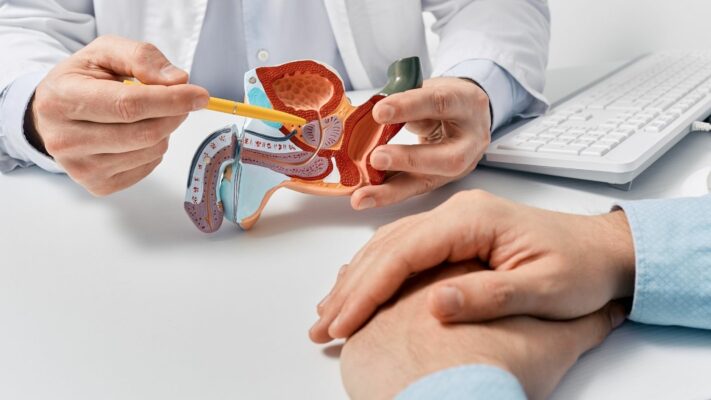
Prostate Problems: Which Doctor or Specialist Should You See?
The medical specialty that men experiencing [...]
Read More
13
Jul
Jul

Premature Ejaculation: Which Doctor Should You See for Treatment?
The first and primary medical specialty [...]
Read More
13
Jul
Jul