
 Türkçe
 Deutsch
 العربية

Percutaneous nephrolithotomy (PNL) is a minimally invasive surgical method that enables large and complex stones in the kidney to be broken and removed through a small access path through the skin. It is performed under general anesthesia and offers a high success rate especially for stones larger than 2 cm.
The procedure of how percutaneous nephrolithotomy surgery is performed begins with the creation of a thin channel from the patient’s back to the kidney, and the stones are reached באמצעות a nephroscope. The stones are broken with ultrasonic or laser devices and removed with special equipment, thus reducing the need for open surgery.
The criteria for to whom PNL surgery is applied include large kidney stones, hard-structured stones, and cases that do not respond to other treatments. It is also preferred in patients with anatomical abnormalities or recurrent stone formation, and appropriate patient selection significantly increases the success rate.
The recovery process after percutaneous nephrolithotomy is generally short, and patients are discharged within a few days. Mild pain, blood in the urine, and the use of a temporary drainage tube may be seen. Follow-up is important against the risk of infection and bleeding, and regular check-ups are recommended.
| What You Need to Know | Information |
| Definition of the Surgery | Percutaneous Nephrolithotomy (PNL) is a minimally invasive surgical method used in the treatment of large, hard, or complex stones located in the kidney. It is based on entering the kidney through a small incision through the skin and breaking and removing the stones. |
| In Which Cases It Is Applied | It is generally preferred for kidney stones larger than 2 cm, stones that do not respond to shock wave therapy (ESWL), staghorn stones, large stones located in the lower calyx, and in the presence of multiple stones. |
| Preoperative Evaluation | Blood tests, urinalysis and culture, kidney function tests, and imaging methods (ultrasonography, computed tomography) are performed. In the presence of infection, antibiotic treatment is planned before surgery. |
| Type of Anesthesia | It is generally performed under general anesthesia. Rarely, spinal anesthesia may be preferred in suitable patients. |
| Surgical Technique | The patient is usually placed in the prone or supine position. An incision of approximately 1 cm is made through the skin. The kidney is entered with a special needle, the tract is dilated, and the stone is visualized with a device called a nephroscope. The stones are broken and removed using ultrasonic, pneumatic, or laser energy sources. |
| Operation Duration | It usually lasts between 1–3 hours depending on the size, number, and location of the stone. |
| Hospital Stay | It usually varies between 2–4 days. The duration may be prolonged depending on the patient’s general condition and whether complications develop. |
| Postoperative Process | A nephrostomy tube may be temporarily placed in the kidney. This tube provides urine drainage and is usually removed within a few days. In some patients, a DJ stent may be placed. |
| Advantages | It provides a high success rate in large and complex kidney stones. Compared to open surgery, it offers a smaller incision, less pain, and a shorter recovery period. |
| Possible Risks and Complications | Bleeding, infection, fever, injury to surrounding organs (intestine, spleen, lung), urine leakage, residual stones, and rarely the need for blood transfusion may be seen. |
| Success Rate | Although it depends on the characteristics of the stone, a stone-free state is generally achieved at a rate of 75–95% in large kidney stones. Additional interventions may be planned if necessary. |
| Recovery Process | Most patients can return to their normal daily activities within a few weeks. Heavy physical activities should be avoided for a certain period according to the doctor’s recommendation. |
| To Whom It Is Not Applied | It should be carefully evaluated in patients with uncontrolled bleeding disorders, in the presence of active and untreated urinary tract infection, and in individuals who carry a high risk for general anesthesia. |
| Alternative Treatment Methods | Shock wave lithotripsy (ESWL), ureterorenoscopy (URS), retrograde intrarenal surgery (RIRS), and rarely open surgery are among the alternative treatment options. |

Prof. Dr. Berkan Reşorlu
Urology Specialist
Urology Specialist
Prof. Dr. Berkan Reşorlu, born in Ankara in 1979, completed his medical education at Ankara University between 1997-2003 and his urology specialization at Ankara University Faculty of Medicine between 2003-2008. Academically, he received the title of associate professor in 2013 and professor in 2022.
He has carried out extensive series studies in the field of endoscopic stone surgery, introduced surgeries applied to children under 1 year of age into the literature, and gained international recognition with his stone scoring system and experimental surgical models.
What Is Urinary System Stone Disease and Why Should It Be Taken Seriously Before PNL?
Kidney and urinary tract stones are a very painful and common health problem that seriously reduces the quality of life. The kidneys, which are the main filtration system of the human body, remove waste substances while cleaning the blood through urine. However, sometimes the concentration of minerals such as calcium, oxalate, and uric acid in the urine increases, or the substances that keep these minerals dissolved are not sufficient. In this case, the minerals gradually precipitate, crystallize, and combine to form hard stone structures. This process, which begins in the form of small grains of sand, can grow over time and turn into stones that fill the internal cavities of the kidney.
These stones not only cause severe, writhing pain; they also silently threaten the health of the kidney. Untreated kidney stones cause an increase in pressure inside the kidney by obstructing the flow of urine from the kidney downward toward the bladder. The pooling of urine inside leads to swelling of the kidney and compression of its tissue. In addition, this stagnant urine creates a perfect breeding ground for bacteria, paving the way for recurrent, persistent, and sometimes life-threatening serious kidney infections that can spread to the blood. As the process continues, permanent damage occurs in the kidney tissue, and eventually the kidney may completely lose its filtering function and fail. For this reason, when kidney stones are diagnosed, it is of vital importance to intervene without delay with a reliable method that will protect the kidney and permanently clear the stones.
In Which Situations Is Percutaneous Nephrolithotomy (PNL) a Preferred Method?
There is no single correct method in the treatment of kidney stones. The size of the stone, its location within the kidney, its hardness, and the patient’s anatomical structure are the most important factors in the treatment decision. Although there are various methods today such as breaking the stone from outside with sound waves or laser fragmentation by entering through the urinary tract, Percutaneous Nephrolithotomy stands out as the most ideal and definitive option in some special situations. Especially in scenarios with large and hard stones where other methods may be insufficient or force the patient to undergo surgery repeatedly, PNL offers high success rates.
The main situations in which this surgery is preferred are as follows:
- Large kidney stones
- Stones located in the lower part of the kidney
- Hard stones that cannot be broken with sound waves
- Stones that cannot be reached through the urinary tract
- Complex staghorn stones
- Kidney stones with congenital anatomical differences
- Patients with narrowing in the urinary tract
- Patients with multiple stones
Why Is PNL the First Choice in Staghorn Stones?
Giant stones with a branched structure resembling coral reefs that fill all or a very large part of the cavities inside the kidney like a mold are called staghorn stones. These stones usually grow very rapidly in connection with urinary tract infections and are extremely dangerous for the kidney. Over time, they silently thin the kidney tissue and consume the kidney from the inside.
Trying to break such a large stone burden by applying sound waves from outside or entering with a laser through the very thin urinary tract is almost impossible. Even if these methods are attempted, complete clearance of the stone may take months, the patient may have to undergo anesthesia repeatedly, and life-threatening infections may develop as a result of broken stone fragments blocking the urinary tract. In contrast, the PNL method provides wide access directly into the kidney through a small tunnel opened from the back. This direct access makes it possible to fragment and vacuum these giant stones and clear them completely in a single session at a high rate.
How Does the Evaluation and Preparation Process Before PNL Surgery Proceed?
The most important step of a successful, safe, and trouble-free surgery is the detailed and meticulous evaluation of the patient before the operation. Before the decision for surgery is made, imaging methods are used in order to see the exact location, size, relation to surrounding organs, and hardness of the stone in three dimensions. This radiological mapping forms the basis of the surgical strategy; it is used to decide where the tunnel will be opened and which instruments will be used.
In addition, the patient’s general health condition, blood values, and risk of infection are examined in detail. Since the kidney is a highly vascular organ, minimizing the risk of bleeding during surgery is a vital issue. For this reason, patients who use blood thinners or anticoagulant drugs need to stop these medications a certain period before surgery or switch to shorter-acting alternative treatments under physician supervision. Also, since the presence of an active infection in the urinary tract can cause microbes to enter the bloodstream during surgery, it is ensured that the urine is completely clean before surgery.
The basic tests that need to be performed before surgery are as follows:
- Computed tomography
- Ultrasonography
- Complete blood count
- Bleeding tests
- Coagulation tests
- Complete urinalysis
- Urine culture
- Kidney function tests
- Liver function tests
- Electrocardiography
Contact us now to get detailed information about treatments and to make an appointment!
What Positions Are Given to the Patient During the PNL Operation?
How the patient lies on the operating table during surgery is a very important detail that directly affects the success and safety of the procedure. This position both provides ease of access to the kidney and allows the anesthesiologist to comfortably control the patient’s respiratory and circulatory systems. Traditionally, for many years the most commonly applied method has been the prone position, in which the patient is laid face down. The patient is first anesthetized in the supine position and then carefully turned over by the operating room team into the prone position. This posture provides a very wide working area for access to the kidney from the back and allows easier access to the avascular regions of the kidney.
However, turning the patient face down while under general anesthesia is a time-consuming procedure that requires attention and may create some physical pressures on the body. Especially in patients who are severely overweight, have serious heart disease, or have low respiratory capacity, lying face down may be risky due to the burden placed on the rib cage. In order to overcome these difficulties and increase patient comfort, new operative positions have been developed in modern surgery.
What Are the Advantages of the Supine PNL Method?
With developing surgical techniques and technological advances, today PNL surgeries performed in the position in which the patient lies completely on their back and their legs are slightly opened to the side with special support have begun to be preferred more and more. This modern approach offers very important benefits especially in terms of patient safety and comfort compared to traditional methods. There is no need to turn the patient again after anesthesia, which both saves time and reduces risks.
The main advantages provided by this method are as follows:
- Higher anesthesia safety
- Shorter operation time
- Less pressure on the airway
- Stability in the heart and circulatory system
- Reduced risk of neck and spine injury
- The possibility of simultaneous intervention through the urinary tract
- Reduced risk of bowel injury
- The surgeon can work more ergonomically while sitting
How Is Safe Access to the Kidney Achieved During PNL?
The most critical and skill-demanding stage of closed kidney stone surgery is creating a thin tunnel extending from the skin on the back to the stone inside the kidney with perfect accuracy. Opening this tunnel at the correct angle and without damaging blood vessels is essential both for the easy clearance of the stone and for preventing possible bleeding. Since it is not possible to do this procedure blindly from the outside, high-tech imaging devices that show the inside of the kidney live are used during surgery.
The most commonly used method is low-dose X-ray devices called fluoroscopy. Thanks to a special contrast substance given into the urinary tract, the internal chambers of the kidney appear clearly on the screen like tree branches. The stone is reached over this map with a thin needle. Under the guidance of a wire passed through the needle, this path is gradually widened millimetrically and a sheath is placed through which surgical cameras can pass. Another method is radiation-free ultrasonography guidance. The location of the kidney and the surrounding organs is seen live with sound waves. Especially in pregnant women, children, and patients who do not want to be exposed to radiation, the use of ultrasound is an extremely safe and modern option.
What Does Miniaturization Mean in PNL Surgical Equipment?
With the development of medical technologies, the size of interventions performed on the human body is getting smaller every day. In the past, the tunnel opened from the back and the sheaths placed into the kidney in closed kidney stone surgeries were relatively thicker. However, today a development called miniaturization has taken place. The aim is to minimize the trauma to the kidney tissue, the risk of bleeding, and postoperative pain by reducing as much as possible the diameter of the tubes placed into the kidney and the optical cameras passing through them.
As the diameter of the instruments decreases, the need to place a drainage tube in the patient’s back after surgery also decreases. In this technique called tubeless PNL, patients go through the postoperative period much more comfortably.
The methods applied according to equipment sizes are as follows:
- Standard PNL
- Mini PNL
- Ultra mini PNL
- Micro PNL
With Which Technologies Are the Stones Inside the Kidney Broken in the PNL Procedure?
After entering the kidney through a very thin tunnel with very high-resolution cameras and clearly visualizing the stone by illuminating it, it is necessary to fragment this stone in order to remove it. Different energy sources have been developed from past to present to break the stone, and these technologies are carefully selected during surgery according to the structure of the stone. Some hard stones require mechanical impacts, while others are vaporized with laser.
Pneumatic lithotripters work with mechanical impacts like a small drill or air gun and can easily break very hard stones. Ultrasonic lithotripters break the stone with the power of sound waves while at the same time aspirating the resulting sand and dust with the help of an integrated vacuum system, which is especially useful for the rapid clearance of large stones. Today’s most advanced stone fragmentation technology is holmium laser systems. The intense laser beam delivered through a flexible and very thin glass fiber melts the stone and turns it almost into sand and dust regardless of the type of stone. Laser technology provides excellent breaking power without damaging the surrounding tissues, especially in procedures performed with very small-diameter cameras.
Contact us now to get detailed information about treatments and to make an appointment!
What Are the Possible Risks and Complications of PNL Surgery?
Every surgical intervention, no matter how modern and closed it is, has some unique risks by the nature of human biology. Closed kidney stone surgery also carries the potential for various complications because it is performed on a vital and highly vascular organ such as the kidney. However, when performed in experienced hands, with modern technological equipment and the right patient selection, these risks are at an extremely manageable level and are overcome without harming the patient. Even when unexpected situations develop, modern medicine has alternative treatments to solve these problems.
The possible risks that may be seen related to this operation are as follows:
- Bleeding in the urine
- Postoperative fever
- Chills and shivering
- Urinary tract infection
- Injury to surrounding organs
- Failure to fully reach the stone
- Stone fragments remaining inside
- Urine leakage
What Are the Factors Affecting the Success of PNL Surgery?
The ultimate goal in closed kidney stone surgeries is to clear the patient’s kidney completely of stones without leaving any fragments behind. However, many variables affect this complete success rate. The total volume of the stone, how much it is spread into different chambers within the kidney, how hard its chemical structure is, and the congenital anatomical shape of the kidney are among the foremost of these factors. In addition, if the patient has previously undergone open kidney surgery, this may cause adhesions around the kidney, making access to the kidney technically more difficult. Nevertheless, thanks to today’s flexible instruments and laser technologies, even stones that were once considered unreachable can now be reached and the surgery can be completed with high success and safety.
How Do Hospitalization and Recovery Progress After PNL Surgery?
After the operation is completed, the patient is awakened and, after being observed for a short time in the recovery room, is taken to their room. Generally, the length of hospital stay for patients who undergo this surgery varies between one and two days depending on their health condition. This process is passed quite calmly with supportive treatments so that the body can eliminate the effects of anesthesia and the kidney can adapt to the new condition. The patient’s pain is kept under control with special medications and fluid support is provided.
Immediately after surgery, there may be a catheter in the patient’s urinary tract and a thin tube at the entry point on the back to accelerate healing. These tubes allow fluids that may accumulate inside and possible small bleedings to be easily drained, thereby relieving the kidney. The urinary catheter is usually removed painlessly on the morning after surgery. When the fluid coming from the tube on the back becomes completely clear, this tube is also removed, and that tiny entry site on the back is closed with a simple dressing and left to heal. The patient, who passes the postoperative period comfortably, is sent home with detailed recommendations.
What Should Be Considered During the Recovery Process at Home After PNL?
When discharged from the hospital, patients can generally return comfortably to desk jobs or normal-paced social life within seven to ten days. That small wound on the back closes and heals completely within a few days. However, the kidney’s internal healing takes a little longer. For this reason, for two weeks after discharge, heavy lifting, sports that excessively strain the body, and intense physical activities must definitely be avoided. Resting well and giving the body time is the golden rule of recovery.
In addition, it is completely normal during this period to occasionally see mild pinkish discoloration in the urine, and this should not cause concern. However, there are also some symptoms that must be monitored carefully during the time spent at home and that require urgent intervention:
The situations in which it is necessary to apply to the hospital are as follows:
- High fever
- Shivering attacks
- Severe flank pain
- Heavy fresh blood in the urine
- Large blood clots in the urine
- Sudden cessation of urine output
- Severe nausea
- Uncontrollable vomiting
How Can the Recurrence of Kidney Stones After PNL Be Prevented?
Unfortunately, complete clearance of all stones from the kidney with a successful surgery does not mean that the patient will never form stones again in the coming years. Kidney stone disease is actually a chronic metabolic problem related to the chemical balance of the body and is quite prone to recurrence. Surgery is a mechanical intervention that saves the kidney and clears the stones; however, it cannot resolve the root cause of the disease on its own. For this reason, surgery should be seen not as a finish line but as a new starting point for a stone-free and healthy life. In order for patients not to experience the same pains again, for kidney functions to be preserved, and for there not to be a need for another surgical intervention in the future, it is absolutely necessary to perform a detailed metabolic evaluation in the postoperative period. The removed stone is sent to the laboratory to determine its type, and by analyzing the patient’s blood and 24-hour collected urine, the cause of stone formation is identified and a personalized preventive strategy is drawn accordingly.
How Should the Diet Be Organized to Prevent Stone Formation After PNL?
The most powerful way to permanently prevent kidney stone formation is conscious and lasting changes in daily eating and lifestyle habits. Although drug treatments may be necessary in some cases, the chance of success is low unless the dietary pattern is placed on solid foundations. The amount of fluid taken into the body, salt consumption, and the content of foods directly affect the filtering burden of the kidney. There are also many misconceptions in society about this issue. For example, a person who forms calcium stones completely removing milk and cheese products from their life can not only lead to bone loss but may also pave the way for even more stone formation by disrupting intestinal balance. The important thing is not to prohibit but to balance.
The basic dietary and lifestyle rules that should be applied in daily life are as follows:
- Plenty of water consumption
- Monitoring light-colored urine
- Restriction of salt use
- Avoidance of processed foods
- Balancing animal protein
- Balanced calcium intake
- Plenty of vegetable consumption
- Plenty of fruit consumption
- Stopping sugary carbonated drinks
- Regular physical activity
Blog Posts

Genital Warts: Which Doctor or Department Should You See?
When genital warts are noticed, the [...]
Read More
13
Jul
Jul

Urinary Tract Infection (UTI): Which Doctor or Department Should You Visit?
The department patients experiencing urinary tract [...]
Read More
13
Jul
Jul
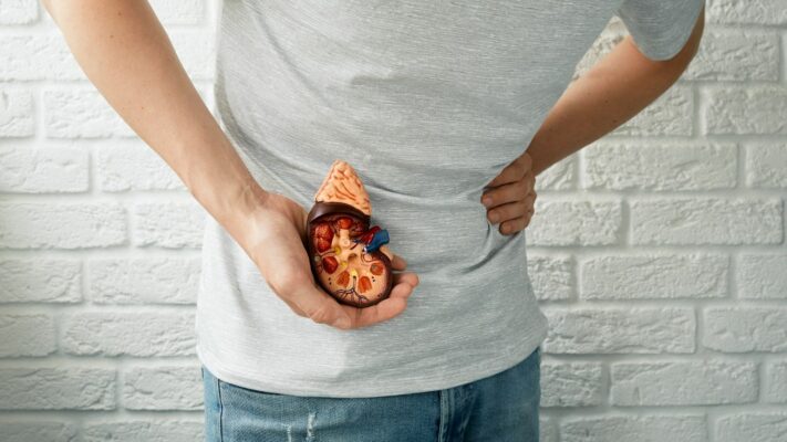
Kidney Stones: Which Doctor or Specialist Should You See?
The primary specialty that patients with [...]
Read More
13
Jul
Jul
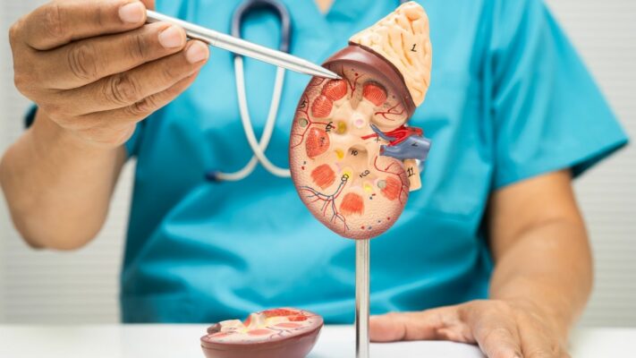
Kidney Diseases: Which Doctor or Department Should You See?
The primary medical specialty that patients [...]
Read More
13
Jul
Jul

Sexually Transmitted Diseases (STDs): Which Doctor Should You See?
The answer to the question of [...]
Read More
13
Jul
Jul
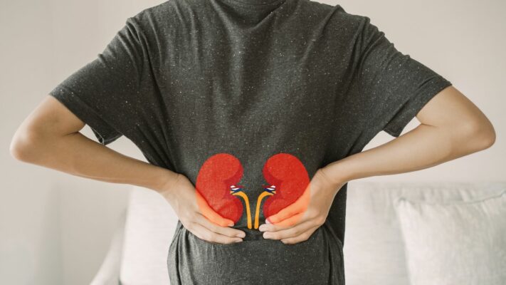
Kidney Cysts: Which Doctor or Department Should You See?
The primary medical specialties that patients [...]
Read More
13
Jul
Jul
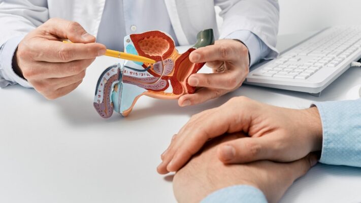
Prostate Problems: Which Doctor or Specialist Should You See?
The medical specialty that men experiencing [...]
Read More
13
Jul
Jul

Premature Ejaculation: Which Doctor Should You See for Treatment?
The first and primary medical specialty [...]
Read More
13
Jul
Jul