
 Türkçe
 Deutsch
 العربية

Adrenal tumor surgery is an effective treatment method that provides the surgical removal of benign or malignant masses developing in the adrenal gland, aiming to regulate hormonal balance and eliminate the tumor burden.
In the process of how adrenal tumor surgery is performed, laparoscopic or open surgical techniques are preferred, and the size of the tumor, its location, and suspicion of malignancy are decisive; minimally invasive methods offer faster recovery and lower complication rates.
The recovery period after adrenal tumor surgery varies depending on the patient’s general health condition and the surgical technique applied; hormonal balance monitoring, infection control, and regular clinical follow-up play a critical role in preventing complications.
Among the risks and complications of adrenal tumor surgery are bleeding, infection, hormonal imbalances, and rarely the possibility of damage to surrounding tissues; therefore, comprehensive preoperative evaluation and the selection of an experienced surgical team are important.
| What You Need to Know | Information |
| Definition of the Surgery | Adrenal tumor surgery is the procedure of surgically removing benign (adenoma, pheochromocytoma, etc.) or malignant (adrenocortical carcinoma, metastasis) tumors developing in the adrenal gland. |
| Function of the Adrenal Glands | The adrenal glands produce vital hormones such as cortisol, aldosterone, adrenaline, and noradrenaline. These hormones affect metabolism, blood pressure, stress response, and electrolyte balance. |
| Surgical Indications | Hormone-secreting tumors (Cushing syndrome, Conn syndrome, pheochromocytoma), masses larger than 4 cm, suspicion of malignancy, tumors showing rapid growth, or those causing symptoms may require surgery. |
| Types of Surgery | There are two main surgical methods: laparoscopic adrenalectomy (closed method) and open adrenalectomy. The size of the tumor, its spread, and the patient’s general condition determine the method. |
| Laparoscopic Method | It is performed using a camera and special surgical instruments through small incisions. It provides less pain, a shorter hospital stay, and faster recovery. |
| Open Surgery | It is preferred in large tumors or tumors that have spread to surrounding tissues. It requires a wider incision and the recovery period is longer. |
| Preoperative Preparation | Hormone levels are examined in detail. Especially in pheochromocytoma, drug treatment is applied before surgery to balance blood pressure. The tumor is evaluated with imaging methods (CT, MRI). |
| Duration of Surgery | It varies between an average of 1 and 3 hours. The duration may be longer depending on the size of the tumor and the surgical method. |
| Anesthesia | It is performed under general anesthesia. |
| Length of Hospital Stay | It is generally 2–4 days in laparoscopic surgery and may be longer in open surgery. |
| Possible Risks | Bleeding, infection, injury to neighboring organs, hormonal imbalances, fluctuations in blood pressure, and rarely clot formation may be seen. |
| Postoperative Follow-up | Hormone levels are monitored. Temporary or permanent hormone replacement therapy may be started when necessary. |
| Recovery Process | Return to daily activities in closed surgery generally takes 2–4 weeks, while in open surgery it may take 4–6 weeks. |
| Hormone Replacement | If both adrenal glands are removed, cortisol and, if necessary, other hormones must be administered externally for life. |
| Pathology Examination | The removed tumor is examined histopathologically and the definitive diagnosis is determined. This result determines the need for additional treatment. |
| Follow-up Process | In malignant tumors, long-term follow-up with regular imaging and hormone tests is necessary. |

Prof. Dr. Berkan Reşorlu
Urology Specialist
Urology Specialist
Prof. Dr. Berkan Reşorlu, born in Ankara in 1979, completed his medical education at Ankara University between 1997-2003 and his urology specialization at Ankara University Faculty of Medicine between 2003-2008. Academically, he received the title of associate professor in 2013 and professor in 2022.
He has carried out extensive series studies in the field of endoscopic stone surgery, introduced surgeries applied to children under 1 year of age into the literature, and gained international recognition with his stone scoring system and experimental surgical models.
Where are the adrenal glands located in our body and what are their functions in terms of adrenal tumors?
The adrenal glands are small, yellowish organs located at the back of the abdominal cavity, positioned like caps right on the top of both kidneys. Although their size is only a few centimeters and their weight is only a few grams, they function almost like the body’s energy and balance control centers. The structure of these small organs consists of two main parts: an outer shell (cortex) and an inner core (medulla). Adrenal tumors also originate from the cells of these two different layers and cause completely different effects in the body depending on which layer they arise from.
The outer cortical region produces three basic steroid hormones that are critically important for the continuation of life. The first of these is aldosterone, which balances blood pressure by regulating the body’s salt and water ratio. The second is cortisol, which is the body’s greatest shield against stress and regulates the immune system and blood sugar. The third is auxiliary androgen hormones that support sexual characteristics. The inner medullary region is in direct communication with the nervous system. It produces hormones such as adrenaline and noradrenaline, which enter the blood within seconds during moments of fear, excitement, or danger. These hormones accelerate the heartbeat, constrict blood vessels, and coordinate the body’s sudden responses to stress factors within seconds. The smooth functioning of this entire complex system depends on the adrenal glands having a completely healthy cellular structure.
How do adrenal tumors arise and what are their characteristics in terms of being benign or malignant?
Cells in the human body grow, divide, and disappear when the time comes according to a certain order and code. However, sometimes changes in the genetic code of the cells, the cause of which cannot always be known, disrupt this perfect cycle. Instead of dying, the cells begin to divide uncontrollably and accumulate where they are located. When these accumulations occur in the adrenal gland, they form masses. A large majority of these masses are benign growths known medically as adenomas. Benign masses usually grow slowly, do not attack surrounding tissues, and do not tend to spread to other organs. These masses mostly follow a silent course.
On the other hand, although quite rare, malignant cancer types called adrenocortical carcinoma may also develop. Malignant adrenal tumors have a much more aggressive and destructive structure. They tend to grow rapidly and may spread directly beyond the adrenal gland into surrounding tissues such as the kidney, liver, and major vessels. Their risk of reaching distant organs such as the lungs or bones through the blood and lymphatic pathways is also quite high. Therefore, when a mass is detected in the adrenal gland, the main purpose of all the tests and imaging performed is to clearly distinguish whether this mass is an innocent adenoma or a dangerous cluster of cancer cells. The chance of modern treatment is very high in masses detected at an early stage.
Into which groups are adrenal tumors divided according to their hormone production status?
Masses formed in the adrenal gland are distinguished from each other not only by whether they are structurally benign or malignant, but also by their ability to produce hormones. Since the main task of this organ is to constantly send certain chemical messengers to the body, a mass developing here may sometimes begin to function independently like a hormone factory on its own. According to these characteristics, tumors are basically divided into two main categories.
The first group is functional, that is, active hormone-producing masses. These masses secrete excessive amounts of hormones continuously regardless of whether the body needs them or not. These high doses of hormones entering the bloodstream disrupt the body’s metabolic balance and pave the way for serious diseases. For example, the continuous pumping of cortisol into the body deeply affects the patient’s entire quality of life. If left untreated, high hormone levels may lead to permanent organ damage.
The second group is non-functional, that is, silent masses that do not produce hormones. Masses in this group do not produce any chemical secretion and can generally remain in the abdominal cavity for years without causing any symptoms. Most of the time, their presence is not felt at all until they become very large and physically compress surrounding organs. Since they do not chemically affect the body’s system, they do not create complaints.
The main hormones secreted are as follows:
- Cortisol
- Aldosterone
- Adrenaline
- Noradrenaline
Contact us now to get detailed information about treatments and to make an appointment!
What do incidentally detected masses (incidentalomas) mean and are these adrenal tumors dangerous?
We have started to hear the word incidentaloma more and more frequently in the medical world and in daily life. This term means a mass detected completely by chance. When a person goes to the doctor for pain in the gallbladder, a severe cough, suspicion of a herniated disc, kidney stone pain, or a completely general check-up screening, various radiological images are taken. Even if the patient’s main complaint is related to a completely different region or system, a mass larger than one centimeter may catch your eye in the adrenal gland on the computed tomography images taken. This surprising situation is called an adrenal incidentaloma.
The biggest reason why this is encountered so frequently is that modern imaging devices are now sensitive enough to show even millimetric details. Especially with advancing age, the possibility of such harmless masses forming in the adrenal glands increases. Statistics clearly show that approximately two or three out of every one hundred people who have no complaints and appear completely healthy have these masses.
When such a mass is found by chance, the first question that comes to mind is understandably how dangerous the situation is. The good news is this: the overwhelming majority of these incidentally detected masses are completely harmless. They do not produce hormones and do not contain cancer cells. However, human health always requires caution. Therefore, no matter how small or innocent the mass appears, it should definitely not be ignored. It must be examined in detail by a specialist, and whether the mass is truly silent and benign should be confirmed with a series of radiological and biochemical blood tests. After confirmation is made, for most people, being followed up at certain intervals is an extremely sufficient and safe approach.
How do radiological imaging and tomography guide the way in suspected adrenal tumors?
When a mass is noticed in the adrenal gland, advanced radiological imaging methods are the most reliable guides in revealing the identity of the mass. In this detailed process, the first device applied and the one that provides the most information about the cellular structure is the Computed Tomography (CT) device. Tomography creates a cross-sectional and highly detailed physical map of the abdominal region and calculates in detail the exact size, smoothness of the borders, and density of the mass.
In radiology, the density of the mass is precisely measured with a special unit called the Hounsfield Unit (HU). Benign and harmless tumors of the adrenal gland structurally contain a large amount of lipid, that is, fat. The density value of fat tissue on tomography is quite low. Therefore, the higher the amount of fat contained in a mass, the higher the probability that that mass is a benign adenoma. However, sometimes masses may not contain a sufficient amount of fat. In that case, a contrast material is given intravenously to the patient in order to better understand the internal structure of the mass.
The time it takes for this medical dye to enter the vascular network of the tumor and the speed at which it is expelled from the tumor are calculated second by second. This is called the washout protocol in radiology. Benign masses clear themselves by returning this dye they take in back to the bloodstream very quickly. Malignant masses or cancer foci, on the other hand, retain this dye inside themselves for a long time.
How are hormone tests of adrenal gland masses or adrenal tumors performed and what diseases do they cause?
While radiological images clearly show the physical structure of the mass, detailed laboratory tests reveal whether the mass is making a chemical attack on the body. Even if it is millimetric in size, hormone screening in blood and urine must definitely be performed for every detected mass. Because even a small and innocent-looking tumor may pave the way for cardiovascular diseases or severe metabolic disorders by continuously pumping hormones into the body.
Excessive secretion of hormones leads to certain specific disease patterns in the body. In order to detect these diseases early, special blood tests and generally urine analyses collected in a special container for a full twenty-four hours are of great importance. These samples taken from patients are subjected to highly sensitive measurements in laboratories.
The main diseases caused by excessive hormone production are as follows:
- Cushing Syndrome
- Conn Syndrome
- Pheochromocytoma
If the tumor produces an excessive amount of cortisol, a picture of Cushing Syndrome occurs. This condition is diagnosed by monitoring how the blood values change the next morning with a small pill swallowed by the patient before going to bed at night.
The symptoms of Cushing syndrome are as follows:
- Weight gain
- Rounding of the face
- Purple stretch marks on the skin
- Muscle weakness
- Bone loss
If the mass produces an excessive amount of the hormone aldosterone, this special picture is called Conn Syndrome. In this disease, while the potassium level in the blood drops to critically low levels, blood pressure becomes resistant even to multiple medications given and rises to dangerous levels.
The third and one of the most exhausting conditions for the body is Pheochromocytoma. These tumors keep the body in constant alarm as if it were continuously in a state of war by secreting adrenaline and noradrenaline continuously.
The symptoms during a pheochromocytoma attack are as follows:
- Severe palpitations
- Excessive sweating
- Fear of death
- Sudden headache
- Very high blood pressure
Contact us now to get detailed information about treatments and to make an appointment!
For adrenal tumors of what size and characteristics is the decision for surgery made?
The presence of a mass in the adrenal gland never means that one should immediately lie down on the operating table. The decision for surgical intervention is made with very fine calculations in order to improve the patient’s quality of life and permanently eliminate possible vital risks. Two main red lines are of great importance when deciding whether a mass should be removed surgically. These are the growth rate of the mass and whether it produces hormones.
If detailed tests prove that the tumor is actively producing hormones, no matter if the physical size of the tumor is one centimeter, that mass should be safely removed from the body surgically. Because the problems created by that small mass, such as high blood pressure, stubborn diabetes, or bone loss, cannot be permanently corrected with medications. The only definite solution is the complete removal of that diseased tissue.
If the mass is not producing hormones and is completely silent, then the focus is on size. Masses under four centimeters, if they appear smooth-bordered on imaging, are generally not operated on. Patients are called for check-ups at certain intervals and the size of the mass is closely followed.
When the mass is between four and six centimeters in size, this region is considered a gray zone in terms of decision-making. In masses of this size, the risk of harboring malignant cells is at a certain level. At this stage, taking into account the patient’s general health condition and the suspicious appearance of the mass on imaging, the decision is generally made in favor of surgery.
If the mass is larger than six centimeters, since the risk of cancer is now considered high, waiting provides no benefit to the patient. Since these masses may grow rapidly and damage surrounding tissues, surgical planning should be made without wasting time. Masses in which rapid growth is detected during follow-up must also definitely be operated on.
Why is preoperative preparation of vital importance for adrenal tumors called pheochromocytoma?
Among adrenal gland surgeries, the most careful and most difficult preparation process is the removal of masses that secrete intense adrenaline. These tumors are extremely active by nature. If the patient is taken directly to the operating room and anesthetized without any medical preparation, the tumor may release a massive amount of adrenaline into the body within seconds.
This high dose of adrenaline suddenly entering the blood causes the vessels to constrict suddenly and violently. This may cause the patient’s blood pressure to shoot up to very serious levels, disrupt the heart rhythm, or lead to cerebral hemorrhage. In the past, risk rates were quite high because this body mechanism could not be managed. However, thanks to modern preparation protocols applied today, this risk has been brought under control.
The critical preparation stages for a successful surgery are as follows:
- Starting alpha-blocker medications
- Gradually lowering blood pressure
- Increasing blood volume with fluids
- Adding beta-blocker medications if necessary
About two to three weeks before the surgery, special vasodilating blood pressure medications are given to the patient. The main task of these medications is to physically prevent the vessels from constricting no matter how much adrenaline is secreted from the tumor. When the patient’s blood pressure is brought to the targeted levels with drug treatment, it is understood that the body has now formed a shield against adrenaline storms. After this meticulous preparation is completed, the patient can receive anesthesia and the operation is performed.
What are the modern urological surgical methods currently applied for adrenal tumors?
With the tremendous advancement of medical technology, except for very large cancers, the era of open abdominal surgery in adrenal gland operations has largely been left behind. Today, these operations are performed quite safely almost entirely with laparoscopic and robotic surgery systems known as closed methods.
Laparoscopic surgery is the standard method accepted and widely practiced all over the world. Three or four holes with very small diameters are opened in the patient’s abdominal region. Through these small holes, a camera that illuminates the inside and provides a very clear image, along with long surgical instruments, are inserted. The inside of the abdomen is gently inflated with gas to create a safe working area. With the help of the camera, the other organs are gently pushed aside and the adrenal gland is reached. The mass is separated together with the surrounding tissues, its main vessels are clipped and cut with special clips, and it is removed from the body in a small protective bag.
In recent years, the advanced technological robotic surgery system has started a completely different era in this field. In robotic surgery, instead of standing at the patient’s head, the surgeon sits at a special console in the room and controls the robot’s advanced arms with fingertip devices.
The advantages offered by robotic surgery are as follows:
- Three-dimensional depth perception
- Very high image magnification
- Reducing hand tremor to zero
- Superior maneuverability in a narrow area
- Flexible wrist movements
Especially when working in an area such as the adrenal gland, which is located very deep in the body and right next to major vessels, this high precision provided by the robot increases surgical success. Since there is no large incision scar on the patient’s body, postoperative pain is minimal. Patients start eating in a short time and are usually discharged within a few days and return to their normal lives.
How does the follow-up process work for adrenal tumors after surgery?
In adrenal gland masses, the treatment process is not considered finished when the surgical operation is completed. The patient should also be monitored regularly and with a certain discipline after the operation. This comprehensive process is carried out not by a single specialty; but by the evaluation of a wide team consisting of a urologist, a hormone disease specialist, a radiologist, and a pathologist who examines tissues.
The course of the follow-up process is shaped entirely individually according to the characteristics of the removed tumor. For example, when a tumor that has been secreting high doses of cortisol into the body for a long time is removed, the healthy adrenal gland on the other side cannot become active immediately because it has been at rest for a long time. The moment the mass is removed, the body is deprived of cortisol support. To prevent this imbalance, the patient is temporarily given hormone support pills from outside until the other gland wakes up and begins to function at full capacity on its own.
In benign masses that do not produce hormones and are removed only because they have physically grown, such supportive treatments are not needed. The removed mass is examined in the pathology laboratory down to the finest detail. After it is reported to be definitively benign, the patient is called for follow-up at routine intervals. Generally, for certain years after the operation, the patient’s general health condition is secured with blood tests and medical imaging.
Blog Posts

Genital Warts: Which Doctor or Department Should You See?
When genital warts are noticed, the [...]
Read More
13
Jul
Jul

Urinary Tract Infection (UTI): Which Doctor or Department Should You Visit?
The department patients experiencing urinary tract [...]
Read More
13
Jul
Jul

Kidney Stones: Which Doctor or Specialist Should You See?
The primary specialty that patients with [...]
Read More
13
Jul
Jul

Kidney Diseases: Which Doctor or Department Should You See?
The primary medical specialty that patients [...]
Read More
13
Jul
Jul

Sexually Transmitted Diseases (STDs): Which Doctor Should You See?
The answer to the question of [...]
Read More
13
Jul
Jul

Kidney Cysts: Which Doctor or Department Should You See?
The primary medical specialties that patients [...]
Read More
13
Jul
Jul
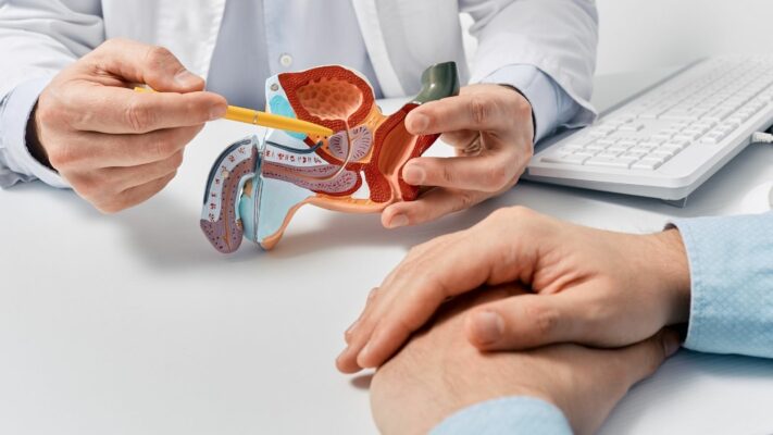
Prostate Problems: Which Doctor or Specialist Should You See?
The medical specialty that men experiencing [...]
Read More
13
Jul
Jul

Premature Ejaculation: Which Doctor Should You See for Treatment?
The first and primary medical specialty [...]
Read More
13
Jul
Jul