Türkçe

Electrolyte testing is a laboratory analysis that measures the levels of essential minerals required for the body’s uninterrupted performance of vital functions such as fluid balance, nerve conduction, and muscle function through blood or urine samples. These biological components, which enable cells to communicate with one another through electrical signals, are the invisible architects of a healthy metabolism. Maintaining the body’s hydration level, regulating blood pressure, and preserving internal balance within ideal ranges all depend directly on these chemical building blocks. Used in a wide range of situations, from routine health screenings to identifying the source of sudden metabolic complaints, this medical examination is one of the most reliable diagnostic methods for assessing organ performance and overall physiological health.
Why Is an Electrolyte Test an Important Indicator of How Our Body Functions?
The human body functions like a massive biological factory in which billions of cells constantly communicate, produce energy, and remove waste products. The key elements that manage this factory’s plumbing and electrical network are electrolytes. Sodium, which is found predominantly in the fluid outside cells, plays a major role in regulating the body’s total water content and balancing blood pressure. Potassium, on the other hand, is found mainly inside cells and helps all muscles, especially the heart muscle, contract and relax in harmony. This difference in the distribution of sodium and potassium creates an electrical current across cell membranes, making nerve transmission possible.
Calcium is indispensable not only for bone health but also for blood clotting and nerve signal transmission. Magnesium functions like motor oil that enables hundreds of different chemical reactions to occur in the body. Chloride generally works together with sodium to support fluid balance, while bicarbonate prevents the acidity of the blood from reaching dangerous levels. The primary minerals measured in this test include:
- Sodium
- Potassium
- Calcium
- Magnesium
- Chloride
- Bicarbonate
Even a slight deviation in the level of any of these minerals can trigger chain reactions throughout the body. A simple feeling of fatigue may actually be a sign of magnesium deficiency, while severe leg cramps may indicate an imbalance of potassium or calcium. Therefore, measuring mineral levels in blood and urine is of great importance whenever there is suspicion that the body’s internal balance has been disrupted.
In Which Situations Is an Electrolyte Test Used in the Diagnosis of Urological Diseases?
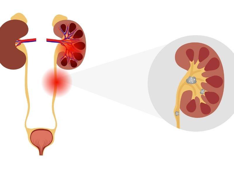
The field of urology examines not only the anatomical structure of the kidneys and urinary tract but also the direct relationship between this system and the body’s chemistry. While filtering the blood, the kidneys produce urine and remove waste products from the body. However, when a condition arises that impairs kidney function—for example, a stone lodged in the urinary tract or an enlarged prostate slowing urine flow—the filtration process begins to deteriorate.
At such times, toxic waste products and minerals that should not accumulate in the bloodstream begin to increase. These tests help determine whether a person’s symptoms are caused solely by a physical obstruction or whether the kidneys’ filtering ability has also been impaired. In addition to obstructions, blood and urine tests are also used to monitor the body’s recovery process following infections that challenge kidney function or major urological surgeries. Some of the symptoms taken into consideration include:
- Severe flank pain
- Inability to urinate
- Body swelling
- Muscle cramps
- Unexplained fatigue
When these symptoms occur, blood and urine analyses provide important clues about the severity of the underlying problem and help shape the patient’s treatment plan.
What Is Investigated With an Electrolyte Test in Recurrent Kidney Stones?
Kidney stones, which are quite common in the general population, are generally considered a warning sign of an underlying disturbance in the body’s chemistry. Individuals who develop stones at a young age, have stones in both kidneys, experience frequent stone episodes, or have only one kidney are considered high-risk patients. In such cases, simply breaking the stone or removing it surgically may not resolve the underlying problem. Unless the metabolic cause is identified, the risk of forming new stones always remains.
To minimize this risk, a comprehensive metabolic evaluation is initiated. The cornerstone of this process is a special analysis in which the patient collects urine continuously over a twenty-four-hour period while maintaining normal daily life and dietary habits. A single spot urine sample may be misleading because it is heavily influenced by daily fluctuations in diet and water intake. Therefore, the entire day’s urine is collected so that the overall average of stone-forming substances and stone-inhibiting protective components can be evaluated. The total urine volume and acidity are also carefully analyzed to develop a personalized dietary and medication plan.
How Are Stone-Forming Substances Evaluated With an Electrolyte Test?
The formation of stones through the aggregation of crystals in the urinary tract is similar to adding too much sugar to a cup of tea, causing the sugar to settle at the bottom. When urine contains too little water and too much solid material, the stone formation process accelerates. The main substances evaluated in urine analysis include:
- Calcium
- Citrate
- Oxalate
- Uric acid
- Sodium
- Cystine
Excessive calcium excretion creates an ideal environment for calcium oxalate stone formation. If high urinary calcium persists despite reducing salt intake, certain diuretic medications that promote calcium reabsorption from the urine back into the bloodstream may be used. Citrate acts as a natural shield by binding to calcium in the urine and preventing crystallization. Low citrate levels increase the risk of stones, and supplements that alkalinize the urine and raise citrate levels may be recommended.
Oxalate is abundant in foods such as spinach, chocolate, tea, and nuts and readily combines with calcium. When oxalate levels are elevated, dietary modifications are recommended to limit these foods. High uric acid levels combined with acidic urine promote uric acid stone formation. In such cases, animal protein intake is reduced and urine acidity is adjusted. Additionally, elevated urinary sodium impairs the kidneys’ ability to reabsorb calcium, making salt restriction advisable. In the rare hereditary condition of cystinuria, abundant water intake and specialized approaches to alter urine chemistry are required.
How Is the Effect of Urinary Tract Obstruction on the Kidneys Determined With an Electrolyte Test?
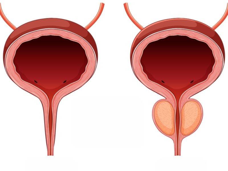
In a healthy urinary system, urine produced by the kidneys flows through narrow ducts into the bladder and is then eliminated from the body. However, when a stone in the urinary tract, benign prostatic enlargement, or a bladder tumor slows or blocks this flow, urine begins to accumulate backward. Like a river blocked by a dam, the accumulating urine creates significant hydrostatic pressure within the kidney tissue. When the delicate filtering units inside the kidneys are exposed to this pressure, their ability to cleanse the blood slows considerably.
As this mechanical obstruction persists, not only waste products such as urea and creatinine, which are normally excreted in the urine, but also potassium begin to accumulate in the bloodstream. Potassium levels exceeding the safe range can lead to serious problems affecting the heart’s electrical system. This condition, which carries risks such as abnormal heart rhythms, often presents with complete inability to urinate and severe abdominal or flank pain. Blood tests quickly measure potassium and kidney function values to determine the extent of damage caused by the obstruction.
What Interventions Are Performed Based on Electrolyte Test Results in Cases of Urinary Tract Obstruction?
In emergencies where kidney filtration has ceased and blood mineral balance has reached dangerously abnormal levels, medication alone is insufficient. The underlying physical obstruction must be relieved promptly to restore urine flow. The primary drainage methods include:
- Urinary catheter placement
- Ureteral stent placement
- Percutaneous nephrostomy
If the obstruction is located at the bladder outlet, liters of retained urine are generally drained using a catheter inserted through the urethra. This procedure rapidly relieves the backward pressure on the kidneys. However, if the obstruction is in the narrow ducts between the kidney and the bladder, special endoscopic instruments are used to place a flexible tube that bypasses the obstruction and allows urine to flow from the kidney into the bladder.
In some cases, the urinary tract may be too severely obstructed to be accessed from below. Under such circumstances, a thin tube is inserted directly into the kidney through the skin of the patient’s back. This method allows urine to drain directly into an external collection bag, relieving pressure on the kidney and allowing blood filtration to resume.
Why Is the Electrolyte Test Repeated Frequently After the Obstruction Has Been Relieved?
Although it may be assumed that the patient will recover quickly once the urinary obstruction has been relieved and urine flow restored, a different and important physiological process begins. Kidneys that have functioned under prolonged high pressure attempt to rapidly eliminate the accumulated waste products from the blood once the obstruction is removed. However, during this period, the kidneys’ ability to concentrate urine and retain water is temporarily impaired.
At this stage, patients begin producing much larger volumes of urine than expected. Daily urine output may reach three or four liters, and very high hourly urine output may also be observed. Similar to opening the floodgates of a dam, this process results in the rapid loss of both water and vital minerals needed by the body. While eliminating accumulated waste products is a positive development, this uncontrolled fluid loss can severely disrupt electrolyte balance. Therefore, laboratory measurements are repeated at frequent intervals to closely monitor the patient’s condition.
How Does an Electrolyte Test Help Protect the Patient During Periods of Excessive Fluid Loss?
If the patient is left without close monitoring during this period of heavy urine output, fluid loss may cause low blood pressure, while the rapid loss of minerals such as sodium and potassium may adversely affect overall body function. Replacing these losses simply by drinking water is generally insufficient. Therefore, the patient is not discharged but remains hospitalized under an intensive fluid monitoring program. The parameters closely monitored during this period include:
- Hourly urine output
- Blood sodium level
- Blood potassium level
- Blood pressure
The patient’s hourly urine volume is measured, and laboratory tests determine the amount of each mineral being lost. Based on these calculations, intravenous fluids containing equivalent amounts of the lost electrolytes are administered. As the kidneys recover over several days and regain their ability to retain water, urine output returns to normal, and intravenous fluid support is gradually reduced and discontinued.
How Is the Risk of Water Intoxication Monitored With an Electrolyte Test During Minimally Invasive Prostate Surgery?
Minimally invasive surgical techniques used to treat benign prostatic enlargement or bladder tumors are performed by entering directly through the urinary tract without making an external incision. Although these procedures, performed with specialized camera-assisted instruments, accelerate recovery, they also carry certain risks. During surgery, irrigation fluid is continuously infused to maintain a clear field of view and flush tissue fragments from the bladder.
As prostate tissue is removed, the surrounding venous vessels become exposed. If the procedure is prolonged or the irrigation fluid is administered under high pressure, this fluid may enter the bloodstream directly through the exposed vessels. Because these irrigation solutions contain no electrolytes, excessive absorption dilutes the blood and rapidly lowers sodium levels. This condition, known as water intoxication, causes water to move from the bloodstream into cells. One of its most serious consequences is the swelling of brain cells due to excessive water uptake. To minimize these risks, blood sodium levels are monitored frequently during and after the procedure.
What Is Done Under the Guidance of an Electrolyte Test When Symptoms of Water Intoxication Develop?
When there is suspicion that irrigation fluid has entered the bloodstream during surgery, physicians must carefully monitor the patient’s neurological and general condition. These operations are generally performed under regional anesthesia, which numbs the lower half of the body. Since the patient remains awake, the earliest signs of water intoxication can be recognized immediately. Some warning symptoms that may be observed during surgery include:
- Confusion
- Nausea
- Vomiting
- Visual disturbances
- Restlessness
- Tremors
If these symptoms develop, bleeding is rapidly controlled, the surgical procedure is terminated, and irrigation is stopped. An urgent blood sample is taken to determine the severity of the sodium decline. If the decrease is mild, potent diuretic medications are administered to eliminate excess water from the bloodstream. However, if sodium levels have fallen to a dangerously low level, intravenous hypertonic saline is administered. This saline treatment must be given slowly and cautiously, as raising sodium too rapidly may cause serious neurological damage.
Why Is Lifelong Electrolyte Testing Necessary After Bladder Removal Surgery?
In the treatment of bladder cancer, the bladder may need to be surgically removed completely to prevent the disease from spreading. After bladder removal, a new system is required to collect and eliminate urine from the kidneys. To create this new urinary pathway, a section of the patient’s small or large intestine is usually removed and reconstructed into a new reservoir or conduit.
The intestine’s new role results in significant long-term physiological changes. Unlike the natural bladder lining, which acts as a barrier that prevents water and minerals from passing through, the intestines are designed to absorb substances into the bloodstream. When urine containing waste products enters this intestinal segment, intestinal cells absorb minerals and acids from the urine and return them to the bloodstream. Since this absorption process directly affects the body’s chemical balance, lifelong monitoring of blood values at regular intervals is essential.
How Are Problems Caused by a New Urinary Tract Constructed From the Intestine Managed With an Electrolyte Test?
When intestinal tissue comes into contact with urine, waste products are absorbed into the bloodstream while substances that regulate blood acidity are secreted into the urine. This reverse exchange causes the blood to become more acidic than normal. This condition, known as acidosis, may lead to persistent fatigue, loss of appetite, weight loss, and easy exhaustion. To keep this imbalance under control, blood gas and mineral measurements are performed regularly. Some supplements that may be required in the long term include:
- Sodium bicarbonate
- Vitamin D
- Vitamin B12
- High-fiber foods
- Antidiarrheal medications
To correct blood acidity, patients are prescribed oral medications that make the blood more alkaline. If acidosis remains undetected for a prolonged period, the body begins drawing calcium from the bones to buffer the acid, creating the foundation for severe osteoporosis in later years. Active vitamin D supplementation is therefore used to help preserve bone health. Additionally, because part of the digestive system has been repositioned, vitamin absorption may become impaired and chronic diarrhea may develop. Regular laboratory testing allows vitamin deficiencies to be detected early and corrected through supplementation, helping patients maintain a healthy life. The success of urological treatments depends not only on surgical procedures but also on maintaining the body’s complex chemical balance over many years with patience and careful monitoring.
Frequently Asked Questions
An electrolyte test is a laboratory test that measures the levels of minerals such as sodium, potassium, chloride, and bicarbonate in the blood. It provides important information about fluid balance, kidney function, nerve conduction, and muscle function.
An electrolyte test may be requested in cases of kidney disease, vomiting, diarrhea, fluid loss, high blood pressure, heart rhythm disorders, muscle weakness, altered consciousness, and while monitoring certain medications.
The test most commonly evaluates sodium (Na), potassium (K), chloride (Cl), and bicarbonate (HCO₃⁻) levels. When necessary, other electrolytes such as calcium, magnesium, and phosphorus may also be examined.
In most cases, fasting is not required for an electrolyte test. However, if the test is planned together with other biochemical analyses, the physician or laboratory may provide specific preparation instructions.
Elevated sodium or potassium levels may be associated with fluid loss, kidney disease, hormonal disorders, certain medications, or metabolic conditions. The results should always be interpreted together with the patient’s clinical findings.
Low electrolyte levels may cause muscle cramps, fatigue, dizziness, palpitations, altered consciousness, nausea, and, in severe cases, seizures. Serious abnormalities may require emergency treatment.
The kidneys play a central role in maintaining electrolyte balance. An electrolyte test helps assess the kidneys’ ability to maintain this balance and is frequently used in the diagnosis and follow-up of kidney diseases.
Insufficient fluid intake, intense exercise, vomiting, diarrhea, medications, kidney and hormonal diseases, and sample collection conditions may all affect electrolyte test results.
The frequency of testing depends on the individual’s health status and the condition being monitored. It may be requested at regular intervals, as recommended by a physician, in patients with kidney disease, heart failure, or those using long-term medications.
An electrolyte test may be requested by specialists in family medicine, internal medicine, nephrology, cardiology, or emergency medicine. The results should always be evaluated by a physician in conjunction with the patient’s clinical condition.

Prof. Dr. Berkan Reşorlu, born in Ankara in 1979, completed his medical education at Ankara University between 1997 and 2003, and his specialization in urology at Ankara University Faculty of Medicine between 2003 and 2008. Academically, he received the title of associate professor in 2013 and professor in 2022.
He has conducted extensive case series studies in the field of endoscopic stone surgery, contributed surgeries performed on children under the age of 1 to the literature, and has gained international recognition with his stone scoring system and experimental surgical models.