
 Türkçe
 Deutsch
 العربية

Bladder cancer surgery is a surgical treatment method that involves partial or complete removal of the bladder depending on the stage and extent of the tumor. The aim is to completely remove the cancerous tissue and prevent the progression of the disease.
Among the types of bladder cancer surgery are transurethral resection (TUR-M), partial cystectomy, and radical cystectomy; these methods are determined according to the depth of the tumor and the patient’s general health condition. Surgical planning requires multidisciplinary evaluation.
Urinary tract reconstruction options after radical cystectomy include ileal loop, orthotopic neobladder, and continent reservoir; these methods aim to preserve the patient’s quality of life. The selection of the appropriate technique depends on individual factors.
The recovery process after bladder cancer surgery is supported by infection control, regular follow-up, and lifestyle adjustments; careful monitoring is required to prevent early complications and improve long-term oncological outcomes.
| Things You Should Know | Information |
| Definition of the Surgery | Bladder cancer surgery is the procedure of removing malignant tumors developing in the bladder through surgical methods. Different surgical techniques are applied depending on the stage, grade of the tumor, and the patient’s general health condition. |
| Types of Surgery | The most commonly applied methods are Transurethral Bladder Tumor Resection (TUR-MT), Partial Cystectomy (removal of part of the bladder), and Radical Cystectomy (removal of the entire bladder and surrounding tissues). |
| TUR-MT (Closed Surgery) | It is applied in early-stage and superficial bladder cancers. The tumor is scraped by entering through the urethra. It is used for diagnosis and treatment. It is usually performed under general or spinal anesthesia. |
| Partial Cystectomy | It can be applied if the tumor is limited to a specific area of the bladder. Only the cancerous part of the bladder is removed. Proper patient selection is important. |
| Radical Cystectomy | It is preferred in cancers that have invaded the muscle layer or are in advanced stages. In men, the prostate and seminal vesicles; in women, the uterus, ovaries, and part of the vagina may also be removed. |
| Urinary Tract Reconstruction | When the bladder is completely removed, methods such as ileal conduit (creating a pouch from the small intestine), orthotopic neobladder (construction of a new bladder), or continent reservoir are applied to expel urine from the body. |
| Duration of Surgery | It may take approximately between 1–6 hours depending on the method applied. Radical cystectomy may take longer. |
| Hospital Stay Duration | After TUR-MT, it is usually 1–2 days; after radical cystectomy, an average hospital stay of 7–14 days may be required. |
| Preoperative Preparation | Blood tests, imaging methods (CT, MR), and anesthesia evaluation are performed. Quitting smoking and controlling existing diseases are recommended. |
| Possible Risks | Bleeding, infection, urine leakage, intestinal complications, anesthesia-related risks, and blood clotting may occur. There may be a risk of loss of sexual function in radical surgeries. |
| Postoperative Process | Catheter use, drain monitoring, and pain control are provided. The normalization of bowel functions is awaited. The pathology result determines the treatment plan. |
| Need for Additional Treatment | Depending on the stage of the tumor, chemotherapy, immunotherapy, or intravesical treatments (such as BCG) may be required. |
| Recovery Process | The full recovery period varies according to the type of surgery. Recovery is faster after TUR-MT; after radical cystectomy, recovery may take weeks to months. |
| Follow-Up Process | Bladder cancer is a disease with a high risk of recurrence. Long-term follow-up with regular cystoscopy, urine cytology, and imaging is required. |

Prof. Dr. Berkan Reşorlu
Urology Specialist
Urology Specialist
Prof. Dr. Berkan Reşorlu, born in Ankara in 1979, completed his medical education at Ankara University between 1997-2003 and his urology specialization at Ankara University Faculty of Medicine between 2003-2008. Academically, he received the title of associate professor in 2013 and professor in 2022.
He has carried out extensive series studies in the field of endoscopic stone surgery, introduced surgeries applied to children under 1 year of age into the literature, and gained international recognition with his stone scoring system and experimental surgical models.
How Does Bladder Cancer Begin in Our Body?
The bladder is an organ made of flexible muscles that serves as a storage unit where urine filtered from the kidneys accumulates before being expelled from the body. A special layer of cells covers the inner surface of this sac, protecting it from the burning and irritating effects of urine. The problem usually begins exactly in this protective inner layer. The contact of harmful substances in the urine with these cells over many years disrupts the natural structure and genetics of the cells. Damaged cells no longer function normally and begin to multiply uncontrollably by dividing. These defective cells, which somehow manage to escape the body’s defense mechanisms, come together to form masses called tumors. Initially, these masses grow on the inner surface of the bladder with a small cauliflower-like appearance, but if not intervened in time, they take root toward the deeper muscle tissues of the bladder. In later stages, they carry the risk of spreading to completely different parts of the body through lymph vessels or blood. Understanding this process of deterioration in cells is of great importance in understanding the stage of the disease.
The main layers found in the structure of the bladder are as follows:
- Urothelium
- Lamina propria
- Detrusor
- Perivesical fat tissue
What Is the Incidence of Bladder Cancer in Türkiye?
When looking at the health data in our country, it is immediately noticeable that this disease shows a very distinct difference between genders. It stands out as the second most common urological problem in men after prostate diseases. When the change over the years is examined, it has been determined that the incidence of the disease is gradually increasing. Especially since the 1990s, the increase in men has accelerated and the figures have almost doubled. Although these figures appear lower in women compared to men, an upward trend in the number of cases is observed with the widespread use of tobacco products among women.
The geography and city in which one lives also have a great impact on the disease. The figures vary in regions where industrialization is intense, where chemical drugs are frequently used in agriculture, or where tobacco consumption is traditionally high. While high rates are observed in provinces such as Antalya, Eskişehir, and Trabzon, these rates decrease somewhat in cities such as Bursa. The stage at which the disease is diagnosed is decisive on survival. Although women generally notice symptoms later and apply to healthcare institutions at a more advanced stage, their overall recovery rates and response to treatment are largely similar to men.
What Are the Most Important Risk Factors for Bladder Cancer?
By far the biggest factor in the emergence of the disease is tobacco products. The probability of a person who smokes developing this disease is approximately three times higher than someone who has never smoked. Thousands of toxic substances in the inhaled smoke mix into the blood and pass into the urine by being filtered by the kidneys. While this toxic urine remains in the bladder for hours, it exposes the inner surface to a kind of chemical bath. The longer the contact time, the higher the probability of the cells becoming cancerous. However, this risk decreases rapidly when smoking is stopped. While the risk decreases noticeably within the first few years, the likelihood of the disease largely disappears when it is not smoked for many years.
Apart from this, occupational exposure to certain chemicals also creates a serious danger. Chemical substances and aromatic amines used in the paint, textile, leather, and rubber industries can cause the disease years later. Drinking insufficient water during the day is also an indirect risk as it prolongs the time urine stays in the bladder. Consuming plenty of water accelerates urine output and provides a protective effect by constantly washing the bladder.
The main risk factors involved in the development of the disease are as follows:
- Tobacco use
- Industrial chemicals
- Chronic bladder infections
- Untreated bladder stones
- Insufficient fluid intake
What Symptoms Does Bladder Cancer Show?
The disease usually progresses silently in the early stages and does not cause much discomfort. However, the first symptom that causes patients the most panic and brings them to a healthcare institution is the presence of blood in the urine. This condition, medically called painless macroscopic hematuria, is the first sign in the vast majority of cases. The color of the urine can vary from light pink to rust color or dark, clotted red. The most misleading part is that this bleeding may sometimes occur only once and not recur for weeks. This leads to postponing visiting a doctor with the thought that the bleeding has passed, which makes early diagnosis difficult.
Of course, seeing blood in the urine does not always mean cancer. Kidney stones, urinary tract infections, or benign prostate enlargement can also cause similar bleeding. However, if there is clotted bleeding that occurs without pain, the situation must be investigated in detail. As irritation in the bladder wall increases, different urinary complaints may also be added to the picture.
Other symptoms of the disease are as follows:
- Frequent urination
- Burning while urinating
- Feeling of not being able to empty the urine completely
- Feeling of pressure in the groin
Which Tests Are Performed in Case of Suspected Bladder Cancer?
After the patient explains their complaints, a detailed physical evaluation is performed. In addition to manual examination of the abdominal area, special examination techniques are used to understand the condition of the organs in the pelvic region. In this way, clinical preliminary information is obtained about whether there is a possible mass at the base of the bladder or whether the tumor has spread outside the bladder. This manual examination is very valuable in planning the next steps.
On the laboratory side, the first step is a urinalysis. The presence of blood cells or an infection in the urine is checked. More importantly, there is the test called urine cytology. Cancer cells that break off from the bladder wall and pass into the urine are searched under a microscope in the pathology laboratory. This test is similar to understanding the type of tree by looking at the leaves that have fallen from it. It is quite successful especially in detecting aggressive and fast-growing cancer cells. However, it may sometimes miss slow-growing tumors. Therefore, the diagnosis is not based on this test alone and must be supported by imaging methods.
The main examinations used in the diagnostic process are as follows:
- Physical examination
- Urinalysis
- Urine cytology
- Ultrasonography
Contact us now to get detailed information about treatments and to make an appointment!
How Is Cystoscopy Used in the Diagnosis of Bladder Cancer?
The method used to prove the definite presence of the disease and known as the gold standard is cystoscopy. This procedure is based on entering through the urinary canal with a thin, long medical instrument with a small camera and light at its tip and examining the inside of the bladder live on a screen. To minimize the feeling of discomfort, anesthetic and lubricating gels are applied to the urinary canal. In this way, the procedure can be completed quite comfortably and painlessly.
Today, this procedure can be performed quite practically in outpatient conditions. Especially flexible instruments, which have a bendable structure, shorten the procedure time and increase comfort as they adapt to the natural curves of the urinary canal. Lenses with different angles are used to ensure that no corner of the bladder is missed. In recent years, thanks to technological developments, imaging techniques using special color wavelengths in addition to standard light have also been added to cystoscopies. These new-generation light systems make abnormal cell foci that may normally be too flat and small to be noticed visible by illuminating them in a different color. Thus, even the smallest suspicious area on the bladder wall can be detected and mapped with high accuracy.
The technological devices and features used during the cystoscopy procedure are as follows:
- Flexible instruments
- Rigid optical lenses
- Narrow band imaging
- Fluorescent blue light
How Do MR Imaging and the VI-RADS System Affect Bladder Cancer Staging?
After a mass is detected in the bladder, the most critical question is whether this mass remains only on the surface or has progressed to the deeper muscle layer of the bladder. The entire course of treatment is determined by the answer to this question. To understand this, multiparametric magnetic resonance imaging technology is used before surgery. The detailed images obtained are evaluated with a special international scoring system called VI-RADS. This system prevents confusion by ensuring that medical professionals speak the same language.
This system scores the risk of tumor infiltration into the muscle tissue from 1 to 5 by examining the images in detail. Low scores indicate that the tumor is definitely superficial, while high scores indicate that the cancer has entered the muscle tissue, disrupted the wall structure, and may even have reached the surrounding fat tissue. Thanks to this method, highly accurate information about the depth of the cancer is obtained before any surgical intervention is performed. This ensures that the most appropriate treatment strategy is offered to the patient from the very beginning, unnecessary procedures are avoided, or vital interventions are carried out without delay.
How Is TUR-M Surgery Performed for Superficial Bladder Cancer?
When the disease is detected, most cases are still at a superficial stage, meaning they have not yet reached the muscle layer. For patients in this group, the procedure that both establishes a definitive diagnosis and provides the initial treatment is TUR-M surgery performed with a closed method. No incision is made on the body. The bladder is reached by entering through the urinary canal under anesthesia. With the help of special instruments, the tumor tissues inside are shaved down to the healthy tissue at their base and removed. These removed pieces are sent to the laboratory to determine the type and stage of the disease.
The main purpose of the procedure is to leave no visible tumor focus inside and to pathologically confirm that the cancer has not passed into the muscle by taking a reliable sample from the muscle tissue beneath the tumor. Sometimes, if sufficient muscle tissue is not seen in the samples taken in the first surgery or if the tumor type is found to be high-risk, the procedure may need to be repeated a few weeks later. This corrective surgery cleans any microscopic foci that may have remained and prevents the true stage of the disease from being missed. According to the structure of the removed tumor, the patient’s subsequent treatment plan is clarified.
The surgical removal techniques used in modern applications are as follows:
- Standard resection
- En-bloc resection
- Laser vaporization
How Does BCG (Tuberculosis Vaccine) Work in the Treatment of Bladder Cancer?
In patients whose superficial tumor has been removed but who are found to have a high risk of recurrence or progression of the disease, preventive treatments come into play. The most effective method applied at this point is BCG therapy, which is administered directly into the bladder and is actually a weakened tuberculosis vaccine. The aim of this vaccine is not to directly poison cancer cells; on the contrary, it is to initiate a tremendous immune response within the bladder. The body’s own defense soldiers are thus awakened.
When the drug is given into the bladder with the help of a catheter, the bacteria attach to the inner surface. The body’s defense system perceives this as a threat and quickly sends its warrior cells to that area. During this intense immune battle that occurs, microscopic cancer cells trying to hide in the environment are also recognized and destroyed by the immune system. The treatment usually starts a few weeks after surgery and is applied in weekly sessions according to a certain schedule. The patient is asked to move so that the drug remains in the bladder for a certain period and comes into contact with every area.
The normal and temporary conditions that may be seen after BCG treatment are as follows:
- Frequent urination
- Mild burning during urination
- Feeling of fatigue
- Mild fever
Contact us now to get detailed information about treatments and to make an appointment!
What Is Radical Cystectomy for Muscle-Invasive Bladder Cancer?
If the cancer has reached the muscle layer deep within the bladder wall or continues to recur and progress despite protective treatments applied while it is still superficial, a regional and comprehensive surgical intervention is now required. Muscle-invasive bladder cancer has the potential to spread rapidly to other parts of the body. The standard operation performed to eliminate this danger is called radical cystectomy. This surgery aims to completely remove all diseased tissues in the pelvic region from the body and is one of the largest operations in the field of medicine.
During the surgery, not only the bladder is removed; the related organs and connective tissues around it are also cleared. The most vital part of surgical success is the removal of the lymph nodes. Cancer cells use the surrounding lymph pathways as the first stop to spread throughout the body. Therefore, all lymph tissues around the main vessels are meticulously removed. The more extensive and thorough this cleaning is performed, the higher the patient’s chance of completely overcoming the disease and the lower the risk of recurrence.
The structures removed during radical cystectomy surgery according to the patient’s gender are as follows:
- Bladder
- Prostate
- Seminal vesicles
- Uterus
- Ovaries
- Pelvic lymph nodes
Why Is Robotic Surgery Preferred in Bladder Cancer Surgery?
These major surgeries, which in the past were performed by making a large incision in the abdomen, can now be performed much more safely with advanced technology robotic surgery systems. Robotic arms with millimetric precision, controlled by the surgeon from a console, provide perfect maneuverability in deep pelvic regions where the human hand has difficulty reaching. Thanks to three-dimensional and high-resolution cameras, tissues are seen much more clearly and human factors such as hand tremor are completely eliminated.
The greatest comfort provided by robotic surgery is minimizing bleeding in areas with dense vascular networks. Thus, the need for blood transfusion during surgery is significantly reduced. Since there is no large incision, the patient experiences less postoperative pain, and bowel movements return to normal more quickly. The length of hospital stay is shortened and the return to normal daily life is significantly accelerated. All these advantages also positively affect the patient’s morale and motivation.
How Are the Urinary Tracts Reconstructed After Bladder Cancer?
When the bladder is removed from the body, a new system must be established for the safe storage and excretion of urine produced by the kidneys. This process is called urinary diversion. This new arrangement, made using a portion of the intestines, can be designed in different ways depending on the patient’s general health condition and age. Each method has advantages according to the patient’s lifestyle, and the final decision is always made jointly after comprehensive discussions.
In one of the most preferred methods, a short segment of the small intestine is used to connect the urinary tract to the abdominal skin. The patient collects urine in a special odorless external bag. In another method, in order to preserve body integrity, a spherical new pouch is created from the intestine. This new pouch is connected to the natural urinary canal, so the patient can continue to urinate naturally without using a bag. However, since the intestinal muscles cannot contract like a real bladder, it may be necessary to use abdominal muscles while urinating, that is, to strain slightly. With training and time, this new system can be adapted to comfortably.
The new urinary tract options applied after bladder removal are as follows:
- Ileal conduit
- Orthotopic artificial bladder
- Continent cutaneous diversion
What Are Smart Drugs in the Treatment of Advanced Bladder Cancer?
When the disease does not remain confined to the bladder and spreads to other parts of the body, the form of treatment completely changes and systemic drugs that will affect the entire body come into play. Standard chemotherapy methods that have been known for years are still successfully used before or after surgery. However, the real revolution in the field of oncology has occurred with smart drugs and immunotherapies that train the body’s defense cells. These new drugs aim to change the behavior of diseased cells.
Immunotherapies do not directly attack the cancer cell; instead, they remove the protein mask that allows cancer to hide from the immune system. The defense cells now recognize the enemy and begin to destroy it on their own. In some cases, these drugs are given as protection after surgery, while in advanced diseases they are used in combination with new generation drugs that act like targeted smart missiles. In this way, patients’ life expectancy can be significantly extended without the severe side effects of classical chemotherapies such as fatigue, hair loss, or decreased blood values.
Modern medical treatments used in advanced-stage disease are as follows:
- Systemic chemotherapy
- Immunotherapy
- Antibody-drug conjugates
- Targeted molecular drugs
How Should Follow-Up Be After Bladder Cancer Treatment Is Completed?
Even if all surgeries or drug treatments are successfully completed, the process with the disease does not end. Bladder cancer, especially when in superficial forms, may tend to recur months or years later in the same place or in a different area. Therefore, it is of vital importance for patients to attend regular check-ups according to a specific schedule throughout their lives. The frequency and content of the controls are specifically planned according to the initial risk level of the disease.
While annual camera-assisted controls are sufficient after the first year in the low-risk group, cystoscopy procedures are performed much more frequently in the first years in the high-risk group. In addition, tomography scans showing the kidneys and urinary tract and cell screening tests are also included in the process. In patients whose bladder has been removed, detailed imaging is performed to determine whether there are signs of disease elsewhere in the body, and organ functions are closely monitored with blood tests. This strict follow-up ensures that any possible problem is detected at the earliest stage and resolved easily.
The medical examinations used in the follow-up process are as follows:
- Control cystoscopy
- Urine cytology
- Computed tomography
- Kidney function tests
- Vitamin B12 measurement
Blog Posts

Genital Warts: Which Doctor or Department Should You See?
When genital warts are noticed, the [...]
Read More
13
Jul
Jul
Urinary Tract Infection (UTI): Which Doctor or Department Should You Visit?
The department patients experiencing urinary tract [...]
Read More
13
Jul
Jul

Kidney Stones: Which Doctor or Specialist Should You See?
The primary specialty that patients with [...]
Read More
13
Jul
Jul
Kidney Diseases: Which Doctor or Department Should You See?
The primary medical specialty that patients [...]
Read More
13
Jul
Jul
Sexually Transmitted Diseases (STDs): Which Doctor Should You See?
The answer to the question of [...]
Read More
13
Jul
Jul
Kidney Cysts: Which Doctor or Department Should You See?
The primary medical specialties that patients [...]
Read More
13
Jul
Jul
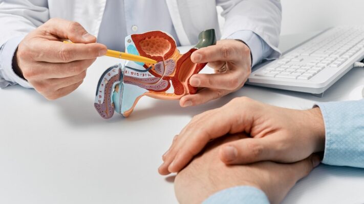
Prostate Problems: Which Doctor or Specialist Should You See?
The medical specialty that men experiencing [...]
Read More
13
Jul
Jul

Premature Ejaculation: Which Doctor Should You See for Treatment?
The first and primary medical specialty [...]
Read More
13
Jul
Jul