
 Türkçe
 Deutsch
 العربية

Kidney stone surgery is a medical intervention that enables the safe removal of stones formed in the kidneys that cannot pass spontaneously, using surgical methods. The size and location of the stone, as well as the patient’s general health condition, are the main criteria in determining the treatment method.
Closed kidney stone surgery methods are modern applications performed using minimally invasive techniques that shorten the patient’s recovery time. Methods such as percutaneous nephrolithotomy and ureteroscopy allow the stone to be broken and removed through small incisions or natural pathways.
The recovery process after kidney stone surgery varies depending on the surgical technique applied and usually allows a quick return to daily life. Drinking plenty of fluids, following the doctor’s recommendations, and not skipping regular check-ups reduce the risk of complications.
Laser kidney stone fragmentation treatment is an effective method that uses high-energy laser technology to break stones into small pieces. This technique is especially preferred for medium and small-sized stones and offers a targeted treatment without damaging surrounding tissues.
| What You Need to Know | Information |
| Definition | Kidney stone surgery is the procedure of removing stones formed in the kidney or urinary tract that cannot pass spontaneously, using surgical or minimally invasive methods. |
| Need for Surgery | Surgical intervention may be required for stones larger than 5–6 mm, causing severe pain, obstructing urine flow, leading to infection, or threatening kidney function. |
| Types of Surgery | Main methods: ESWL (extracorporeal shock wave therapy), Ureterorenoscopy (URS), Percutaneous Nephrolithotomy (PNL), and rarely open/laparoscopic surgery. |
| ESWL | Based on breaking the stone with shock waves sent from outside the body. It is generally applied to small and medium-sized stones. Anesthesia requirement is minimal. |
| Ureterorenoscopy (URS) | A procedure in which a thin endoscope is inserted through the urinary tract and the stone is broken with a laser and removed. It can be applied to ureter and kidney stones. |
| Percutaneous Nephrolithotomy (PNL) | Preferred for large kidney stones. A small incision is made in the back to access the kidney, and the stone is fragmented and removed. |
| Open Surgery | Rarely performed today. It is preferred for very large, complex, or stones that cannot be removed with other methods. |
| Type of Anesthesia | Local, spinal, or general anesthesia may be applied depending on the procedure. |
| Surgery Duration | It usually varies between 30 minutes and 2 hours depending on the method used and the size of the stone. |
| Hospital Stay | ESWL is usually performed on an outpatient basis. After URS and PNL, a hospital stay of 1–3 days may be required. |
| Postoperative Pain | Mild to moderate pain may occur and is controlled with painkillers. |
| Possible Complications | There are risks such as bleeding, infection, urine leakage, ureter injury, and incomplete removal of stone fragments. |
| Recovery Process | Most patients can return to daily activities within a few days. Full recovery time varies depending on the method. |
| Stent Placement | In some patients, a temporary ureteral stent may be placed to ensure urine flow. |
| Risk of Recurrence | Kidney stones may recur. The risk can be reduced with proper diet, fluid intake, and metabolic evaluation. |
| Preventive Measures | Adequate daily water intake, control of salt and oxalate-rich foods, regular follow-up, and medication if necessary are recommended. |

Prof. Dr. Berkan Reşorlu
Urology Specialist
Urology Specialist
Prof. Dr. Berkan Reşorlu, born in Ankara in 1979, completed his medical education at Ankara University between 1997-2003 and his urology specialization at Ankara University Faculty of Medicine between 2003-2008. Academically, he received the title of associate professor in 2013 and professor in 2022.
He has carried out extensive series studies in the field of endoscopic stone surgery, introduced surgeries applied to children under 1 year of age into the literature, and gained international recognition with his stone scoring system and experimental surgical models.
What is a Kidney Stone and How Does It Form in Our Body?
The kidneys, one of the most remarkable filtration systems of the human body, are responsible for continuously cleaning the blood and removing waste substances through urine. Under normal conditions, many minerals and salts such as calcium, oxalate, and uric acid are present in urine in a balanced and completely dissolved form. At the same time, urine also contains important protective substances such as citrate, which prevent these minerals from sticking together and precipitating. However, when water intake is severely insufficient, genetic factors come into play, or unhealthy dietary habits develop, the amount of fluid in the urine decreases and the level of protective substances drops. In this low-fluid environment, dissolved minerals can no longer remain suspended and begin to crystallize.
These microscopic crystals gradually grow like a snowball, harden, and eventually form kidney stones. These stones may remain silent and grow for years without causing any symptoms inside the kidney chambers, or they may remain small and fall into the urinary canal. When a stone enters the urinary canal and suddenly blocks the flow of urine toward the bladder, fluid begins to accumulate in the kidney and the kidney capsule becomes severely stretched. This sudden increase in pressure is the main physical cause of the intense, colicky pain that starts in the lower back and radiates toward the groin, leading patients to seek emergency care.
What Are the Metabolic Factors That Trigger Kidney Stone Formation?
In individuals who experience kidney stones multiple times in their lives, it is well known that simply breaking the stone is not enough and the underlying chemical production error must be identified. At this stage, 24-hour urine collection tests are performed to carefully detect the metabolic and chemical abnormalities that contribute to stone formation. These analyses provide critical information about the stone composition and the patient’s metabolism, forming the basis for personalized preventive treatments to avoid future stones. For example, excessive excretion of calcium absorbed from the intestines or released from bones is one of the most common abnormalities.
The main risk factors evaluated in urine tests are:
- Hypercalciuria
- Hypocitraturia
- Hyperuricosuria
- Hyperoxaluria
These terms have clear clinical meanings. Hypercalciuria refers to excessive calcium in the urine and is the primary cause of calcium oxalate stones. Hypocitraturia indicates a deficiency of citrate, which is a key protective factor preventing crystal formation. Hyperuricosuria refers to high levels of uric acid in the urine and is often associated with excessive consumption of animal protein. In addition, the pH level of urine, which reflects its acidity or alkalinity, is a crucial indicator of metabolic balance. Highly acidic urine creates an ideal environment for uric acid stone formation.
Can a Small Kidney Stone Pass on Its Own?
If imaging methods such as CT or ultrasound reveal that a kidney stone is small, does not cause a dangerous obstruction in the urinary tract, is not associated with infection, and the patient’s pain is controlled with medication, the first preferred approach is usually to wait for the stone to pass naturally. The ureter (the urinary canal between the kidney and bladder) is not a rigid pipe but a flexible structure with contractile ability and certain anatomical narrow areas. Therefore, whether a stone can pass depends entirely on physical conditions. Most stones smaller than 5 millimeters have the potential to pass spontaneously within approximately six weeks. However, as the stone size increases, the likelihood of spontaneous passage decreases significantly.
The main factors determining whether the stone can pass are:
- Size of the stone
- Location
- Width of the canal
- Shape of the stone
The location of the stone in the urinary tract is also highly important. Stones that have moved closer to the bladder have a much higher chance of passing compared to those located near the kidney. Patients are usually monitored for four to six weeks. If the stone does not move, causes significant swelling in the kidney, or the patient continues to experience severe pain, the waiting period is terminated and active surgical treatment becomes necessary.
Are Medications Effective in Kidney Stone Treatment?
The method known among the public as medication-based stone dissolution or passage, medically referred to as Medical Expulsive Therapy, is a highly effective supportive treatment aimed at facilitating the natural passage of stones and improving patient comfort. The lower parts of the urinary tract contain specific receptors in the smooth muscle tissue. Medications used in this treatment, particularly alpha-blockers commonly prescribed for prostate enlargement, target these receptors to relax and slightly widen the urinary canal.
This muscle-relaxing effect does not stop the natural rhythmic contractions (peristalsis) of the ureter; on the contrary, it reduces spasms and allows the stone to slide more easily, almost as if moving through a lubricated tube. As a result, both the frequency and severity of renal colic attacks are significantly reduced. However, medication alone is not sufficient. The patient plays a crucial role in this process. Drinking at least 2.5 to 3 liters of water daily to increase urinary flow and staying physically active to benefit from gravity are essential for accelerating stone passage.
How is Kidney Stone Fragmentation with Sound Waves Performed?
Extracorporeal Shock Wave Lithotripsy (ESWL) is a revolutionary and non-invasive treatment method that has been widely used in kidney stone management for many years. The working principle of this system is quite impressive. High-energy sound waves generated inside the device are transmitted through a water-filled cushion placed in contact with the patient’s body. Since the human body consists largely of water, these waves pass through the skin, muscles, and internal organs without causing damage, much like traveling through water. However, when they hit the dense and hard stone in the kidney, they release their energy. The formation and collapse of microscopic bubbles on the stone surface cause it to gradually fragment into small particles that can be passed through urine. The fact that it usually does not require anesthesia and allows patients to return home immediately after the procedure makes this method very attractive.
Factors that negatively affect success rates include:
- Obesity
- Stone hardness
- Lower pole location
- Large stone volume
The effectiveness of the method varies depending on the characteristics of the stone. It is highly successful for stones smaller than 2 centimeters located in the upper parts of the kidney. However, it is less preferred for stones in the lower pole because gravity makes it difficult for fragments to pass. In addition, increased body mass index reduces the effectiveness of shock waves. After the procedure, patients must maintain high fluid intake to facilitate the passage of fragments.
Contact us now to get detailed information about treatments and to make an appointment!
What is the Closed Laser (URS) Method in Kidney Stone Treatment?
With the development of endoscopic systems and laser technology in urology, Ureterorenoscopy (URS) has become one of the most commonly performed and highly successful surgical methods. This procedure is performed without any external incision on the patient’s body. It is carried out entirely through natural urinary pathways. Using thin, elongated optical instruments equipped with a high-resolution camera and a strong light source, entry is made through the urethra into the bladder and then into the ureter where the stone is located. The stone is visualized clearly on a monitor.
After identifying the stone, a laser fiber is advanced through a small channel within the instrument. The laser energy breaks the stone into small fragments or turns it into fine dust. Remaining fragments can be removed using tiny basket-like instruments. URS is considered the gold standard, especially for stones located in the lower urinary tract that do not pass spontaneously. Thanks to technological advancements, success rates are extremely high. At the end of the procedure, a temporary silicone tube called a Double-J stent is usually placed to prevent swelling and ensure urine flow.
What is Kidney Stone Surgery with Flexible Instruments (RIRC)?
In standard URS procedures, semi-rigid instruments are used, which are effective in the ureter but cannot reach all areas inside the kidney. To overcome this limitation, Retrograde Intrarenal Surgery (RIRC) is used. In this advanced technique, flexible ureteroscopes that can bend up to 270 degrees allow the surgeon to access all chambers of the kidney, including difficult lower pole areas. This method is performed entirely through the natural urinary tract without any external incision, making it one of the most refined surgical techniques available today.
The main principle of this method is not to extract large fragments but to pulverize the stone completely. This technique, known as “dusting,” transforms the stone into fine particles that are easily passed with urine without causing pain. RIRC is especially preferred for stones smaller than 2 centimeters, in obese patients, or in cases where ESWL has failed.
Why is Thulium Laser Technology Important in Kidney Stone Treatment?
For many years, Holmium laser technology has been the primary tool in closed kidney stone surgeries. However, the introduction of Thulium Fiber Laser (TFL) technology has revolutionized urological laser surgery. The working principle of thulium laser is related to its wavelength, which is absorbed by water four times more than Holmium laser. Since kidney stones contain water and are surrounded by fluid, this high absorption allows energy to be delivered directly to the stone.
Innovations provided by Thulium laser include:
- Dusting effect
- Thin fiber usage
- Stable stone targeting
- Faster procedures
These innovations have major clinical advantages. Thinner fibers improve visibility and flexibility of instruments. Unlike older lasers, which push the stone backward (retropulsion), thulium laser keeps the stone stable and gradually breaks it down into fine dust. This reduces surgery time and minimizes complications.
How is PNL Surgery Performed for Large Kidney Stones?
For large kidney stones exceeding 2 centimeters or complex stones filling the kidney like a staghorn, minimally invasive laser techniques may not be sufficient. In such cases, Percutaneous Nephrolithotomy (PNL) is the most effective solution. A small tract is created from the patient’s back into the kidney, approximately the size of a pencil. Through this tract, optical instruments are inserted, and stones are fragmented using powerful devices and removed from the body.
Thanks to technological advancements, Mini-PNL and Micro-PNL techniques now allow the use of much thinner instruments, significantly reducing tissue trauma and bleeding risk. In some modern approaches, drainage tubes are no longer required, allowing patients to be discharged within a day.
What Are the Risks of Kidney Stone Surgery?
Like all surgical procedures, kidney stone surgeries carry certain risks. However, with modern equipment and advanced imaging systems, these risks have been minimized. Most complications are mild and temporary. The most serious risk is infection caused by bacteria entering the bloodstream. Therefore, urine must be sterile before surgery, and infections must be treated beforehand.
Possible mild complications include:
- Fever
- Pain
- Bleeding
- Discomfort
Temporary symptoms such as mild blood in urine or irritation caused by stents are normal and usually resolve after stent removal.
Is Open Surgery Still Performed for Kidney Stones Today?
In the past, kidney stone surgeries involved large incisions and long recovery periods. Today, thanks to technological advancements, open surgery is almost completely obsolete. It is performed only in rare and complex cases where minimally invasive methods are not feasible.
In such cases, laparoscopic surgery is preferred. This technique uses small incisions and specialized instruments, offering faster recovery and less pain compared to traditional open surgery.
What Should We Eat to Prevent New Kidney Stones?
Treatment of kidney stones does not end with surgery. Without lifestyle changes, the risk of recurrence exceeds 50% within five years. The most important dietary rule is reducing salt intake, as excessive sodium increases calcium excretion in urine. Limiting processed foods is essential.
Excessive animal protein intake also increases stone risk by altering urine pH. A balanced diet including plant-based proteins is recommended.
Foods to limit include:
- Spinach
- Beetroot
- Strawberries
- Almonds
- Chocolate
- Tea
Avoid completely eliminating calcium-rich foods, as this may increase oxalate absorption.
Why is Water Consumption So Important in Preventing Kidney Stones?
Drinking plenty of water is the most effective and simplest way to prevent kidney stones. Diluting urine reduces the concentration of minerals and prevents crystallization. The goal is to produce at least 2 to 2.5 liters of urine daily, which requires consuming around 2.5 to 3 liters of fluids.
Urine color is a key indicator: light-colored urine means proper hydration, while dark urine signals risk. Natural beverages like lemonade and orange juice can also help by increasing citrate levels and preventing stone formation.
Blog Posts

Genital Warts: Which Doctor or Department Should You See?
When genital warts are noticed, the [...]
Read More
13
Jul
Jul

Urinary Tract Infection (UTI): Which Doctor or Department Should You Visit?
The department patients experiencing urinary tract [...]
Read More
13
Jul
Jul
Kidney Stones: Which Doctor or Specialist Should You See?
The primary specialty that patients with [...]
Read More
13
Jul
Jul
Kidney Diseases: Which Doctor or Department Should You See?
The primary medical specialty that patients [...]
Read More
13
Jul
Jul
Sexually Transmitted Diseases (STDs): Which Doctor Should You See?
The answer to the question of [...]
Read More
13
Jul
Jul
Kidney Cysts: Which Doctor or Department Should You See?
The primary medical specialties that patients [...]
Read More
13
Jul
Jul
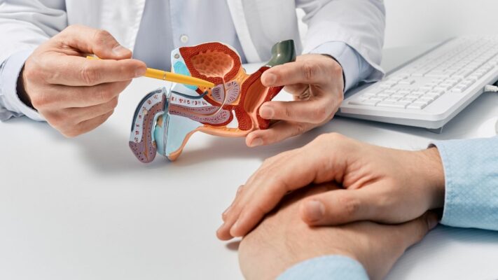
Prostate Problems: Which Doctor or Specialist Should You See?
The medical specialty that men experiencing [...]
Read More
13
Jul
Jul

Premature Ejaculation: Which Doctor Should You See for Treatment?
The first and primary medical specialty [...]
Read More
13
Jul
Jul